. 2011 Feb 1;186(3):1735-46.
doi: 10.4049/jimmunol.1003456. Epub 2010 Dec 27.

Controlling herpes simplex virus-induced ocular inflammatory lesions with the lipid-derived mediator resolvin E1

Affiliations

Controlling herpes simplex virus-induced ocular inflammatory lesions with the lipid-derived mediator resolvin E1

Naveen K Rajasagi et al. J Immunol. .

Abstract

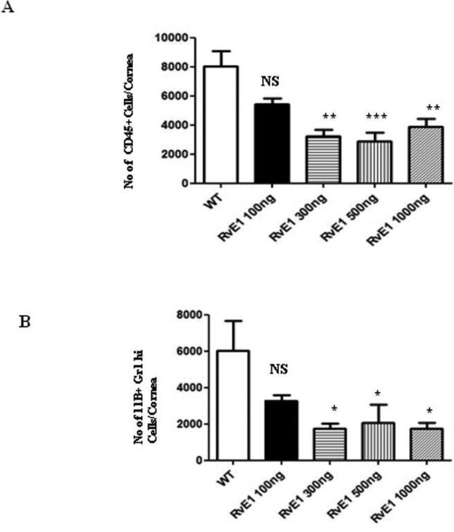
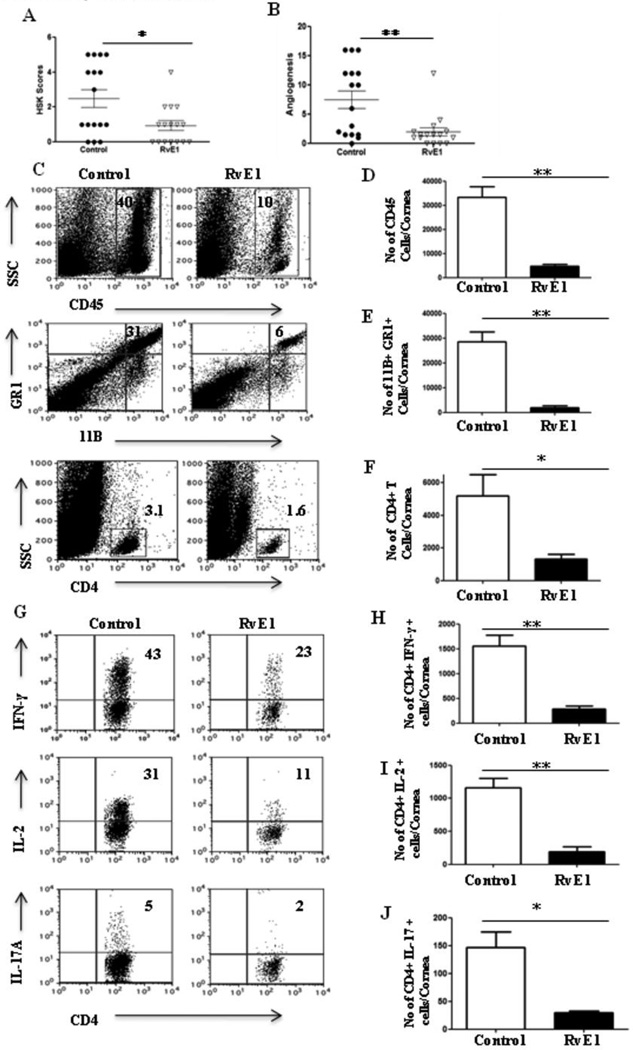
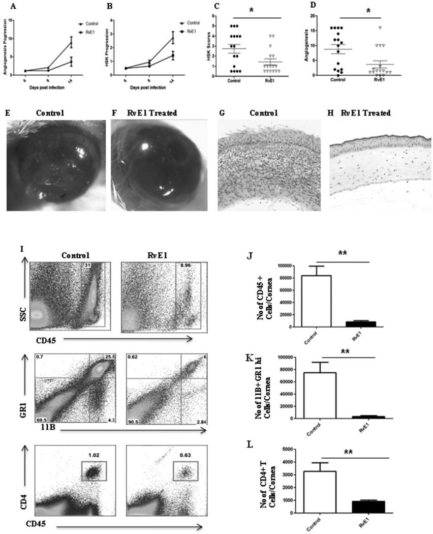
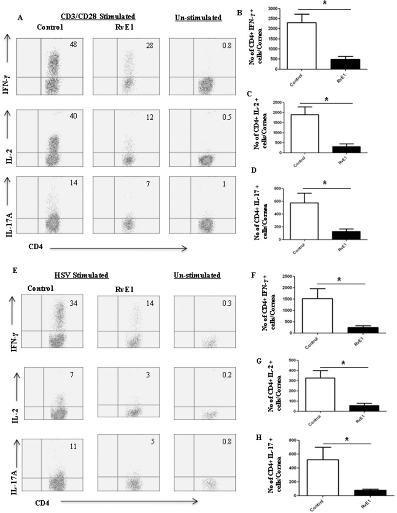
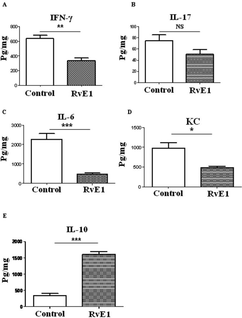
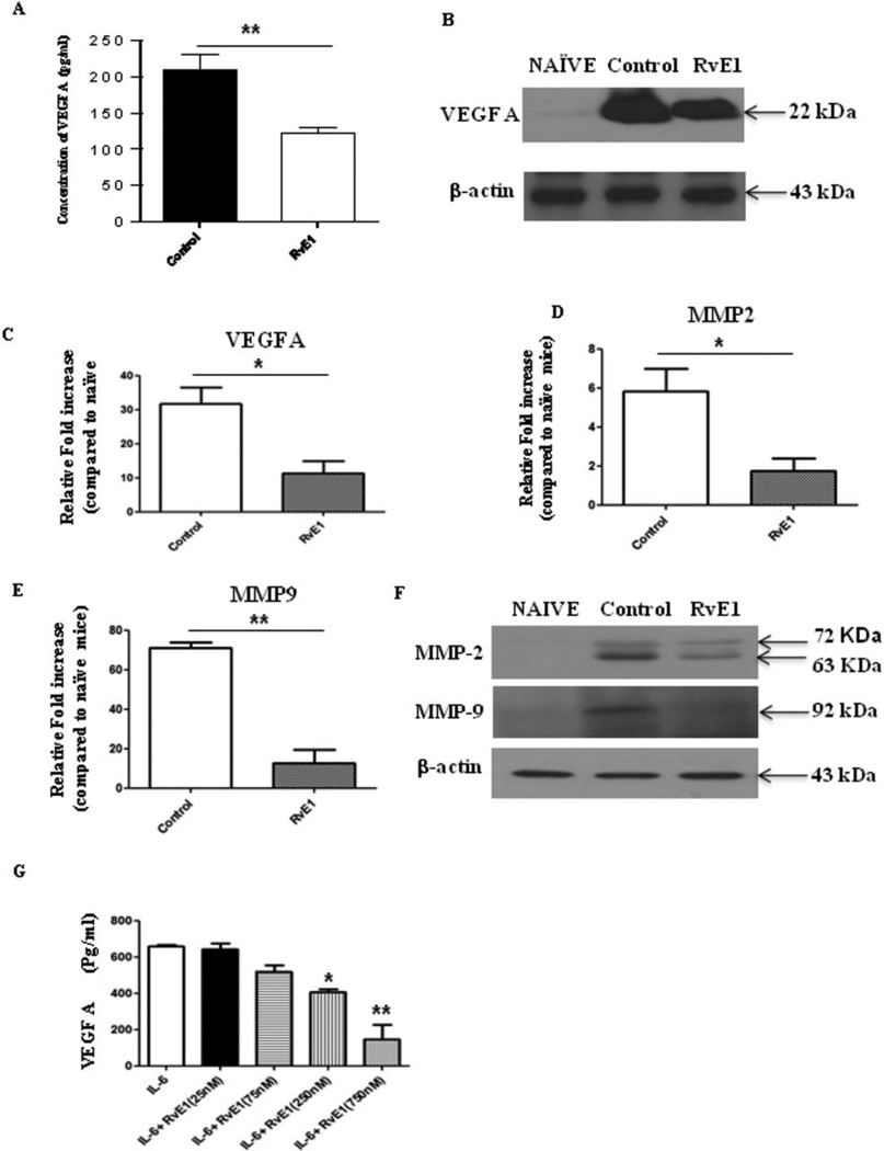
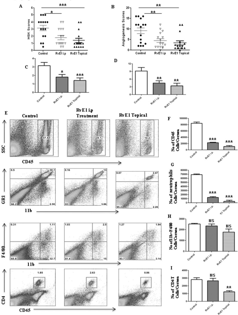
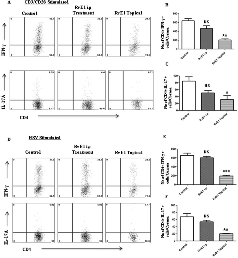
Stromal keratitis (SK) is a chronic immunopathological lesion of the eye caused by HSV-1 infection and a common cause of blindness in humans. The inflammatory lesions are primarily perpetuated by neutrophils with the active participation of CD4(+) T cells. Therefore, targeting these immune cell types represents a potentially valuable form of therapy to reduce the severity of disease. Resolvin E1 (RvE1), an endogenous lipid mediator, was shown to promote resolution in several inflammatory disease models. In the current report, we determined whether RvE1 administration begun at different times after ocular infection of mice with HSV could influence the severity of SK lesions. Treatment with RvE1 significantly reduced the extent of angiogenesis and SK lesions that occurred. RvE1-treated mice had fewer numbers of inflammatory cells that included Th1 and Th17 cells as well as neutrophils in the cornea. The mechanisms by which RvE1 acts appear to be multiple. These included reducing the influx of neutrophils and pathogenic CD4(+) T cells, increasing production of the anti-inflammatory cytokine IL-10, and inhibitory effects on the production of proinflammatory mediators and molecules, such as IL-6, IFN-γ, IL-17, KC, VEGF-A, MMP-2, and MMP-9, that are involved in corneal neovascularization and SK pathogenesis. These findings are, to our knowledge, the first to show that RvE1 treatment could represent a novel approach to control lesion severity in a virally induced immunopathological disease.


Figures

Figure 1
Figure 1. RvE1 reduces cellular infiltration into the cornea of ocularly infected mice in a dose dependent manner
Balb/c mice were ocularly infected with 2×10⁵ of HSV-1 RE. Different doses (100ng, 300ng, 500ng, 1000ng) of RvE1 were applied topically at 18hrs, 24hrs and 36hrs post infection. The animals were sacrificed at 48hrs post infection and pooled corneal samples were analyzed for surface expression of CD45, 11b and Gr1 by flow cytometry. A. Number of leukocytes (CD45+) per cornea and B, Number of neutrophils (11b+ Gr1hi) per cornea at indicated time point is shown. Data shown is from one representative experiment. Experiments were repeated two times. One-way ANOVA was used to calculate the level of significance
Figure 2
Figure 2. Effect of RvE1 started during the pre-clinical phase (day 1 pi) on SK severity and cellular infiltration
Balb/c mice infected with 2×10⁵ PFU of HSV-1 RE were given RvE1 topically twice daily starting from day 1 until day 12. The disease severity and immune parameters were analyzed at day 15. A–B. SK lesion severity and angiogenesis at day 14 are shown. C. Representative histograms show percentage of leukocytes (CD45+), neutrophils (11b+Gr1hi) and CD4+ T cells in the inflamed cornea of control and RvE1 treated animals at day 15. D–F. Average no. of (D) CD45+ cells, (E) 11b+ Gr1hi and (F) CD4+ T cells per cornea at indicated time point are shown. G. Representative plots show percentage of CD4 cells producing IFN-γ, IL-2 or IL-17 following stimulation with CD3/CD28 in the corneas of infected animals. Plots shown were gated on CD4+ T cells. H–J. Average no. of CD4 cells producing (H) IFN-γ, (I) IL-2 and (J) IL-17 in the cornea are shown. Experiments were repeated three times and the level of significance was determined by Student’s t test (unpaired).
Figure 3
Figure 3. Effect of RvE1 started during the clinical phase (day 6 pi) on corneal inflammation and cellular infiltration
Balb/c mice infected with 2×10⁵ PFU of HSV-1 RE were given RvE1 topically twice daily starting from day 6 until day 13. The disease progression and immune parameters were analyzed. A–B. Disease progression of lesions and angiogenesis are shown. C–D. SK lesion severity and angiogenesis at day 14 are shown. E–F. Eye pictures of control and RvE1 treated animals showing lesion severity at 14 days post infection from a representative experiment are shown. Images were captured by stereomicroscopy and an imaging system. Magnification, 20×. E–F. Mice were terminated at day 14 p.i. and eyes were processed for cryosection. H&E staining was performed on 6-µm sections. The figure shows the pictures of the sections taken at 20×, magnification. I. Representative histograms show percentage of leukocytes (CD45+), neutrophils (11b+Gr1hi) and CD4+ T cells in the inflamed cornea of control and RvE1 treated animals at day 15. J–L. Average no. of (J) CD45+ cells, (K) 11b+ Gr1hi and (L) CD4+ T cells per cornea at indicated time point are shown. Experiments were repeated three times and the level of significance was determined by Student’s t test (unpaired).
Figure 4
Figure 4. Effect of RvE1 started during the clinical phase (day 6 pi) on Th1 and Th17 cell infiltration into the inflamed cornea at day 15
Balb/c mice infected with 2×10⁵ PFU of HSV-1 RE were given RvE1 topically twice daily starting from day 6 until day 13.A. Representative plots show percentage of CD4 cells producing IFN-γ, IL-2 or IL-17 following stimulation with CD3/CD28 in the corneas of infected animals. Plots shown were gated on CD4+ T cells. B–D. Average no. of CD4 cells producing (B) IFN-γ, (C) IL-2 and (D) IL-17 in the cornea at day15 are shown. E. Representative plots show percentage of CD4 cells producing IFN-γ, IL-2 or IL-17 following stimulation with HSV-1 in the corneas of infected animals. Plots shown were gated on CD4+ T cells. F–H. Average no. of CD4 cells producing (F) IFN-γ, (G) IL-2 and (H) IL-17 in the cornea at day15 are shown. Experiments were repeated three times and the level of significance was determined by Student’s t test (unpaired).
Figure 5
Figure 5. Effect of RvE1 treatment on cytokine and chemokine production in the corneas of HSV-1 infected animals
Balb/c mice infected with 2×10⁵ PFU of HSV-1 RE were given RvE1 topically twice daily starting from day 6 until day 13. Mice were sacrificed at day 15 and corneal extracts were measured by sandwich ELISA. (A) IFN-γ (B) IL-17 (C) IL-6 (D) KC and (E) IL-10) protein levels in four different pooled corneal samples each consisting of four cornea/group in control and RvE1 treated animals are shown. The level of significance was determined by Student’s t test (unpaired).
Figure 6
Figure 6. Effect of RvE1 treatment on pro-angiogenic factors
Balb/c mice infected with 2×10⁵ PFU of HSV-1 RE were given RvE1 topically twice daily starting from day 6 until day 13. Mice were sacrificed at day 15 and (A) VEGF-A protein levels in four different pooled corneal samples each consisting of four cornea/group in control and RvE1 treated animals were quantified using quantikine sandwich ELISA kit (R&D). B. Immunoblot showing VEGF-A expression in the pooled corneal homogenates from cornea obtained from naïve and HSV-1 infected control and RvE1 treated animals. C–E. Q PCR was used measure the expression of (C) VEGF-A (D) MMP-2 and (E) MMP-9 in pooled corneal samples four cornea/group in control and RvE1 treated animals. The experiments were repeated 3 times. (F) Immunoblot showing MMP-2 and MMP-9 expression in the pooled corneal homogenates from cornea obtained from naive and HSV-1 infected control and RvE1 treated animals. The experiments were repeated two times and the level of significance was determined by Student’s t test (unpaired). G. Effects of RvE1 on VEGF-A production. MK/T-1 cells were stimulated with recombinant murine IL-6 in the presence of various doses of RvE1 for 24 hr period. VEGF-A protein levels in the supernatants were measured using sandwich ELISA. One-way ANOVA was used to calculate the level of significance.
Figure 7
Figure 7. Effect of systemic administration of RvE1 started during the clinical phase (day 6 pi) on corneal inflammation and cellular infiltration
Balb/c mice infected with 2×10⁵ PFU of HSV-1 RE were given RvE1 either intra peritonealy or topically twice daily starting from day 6 until day 13. A–B. SK lesion severity and angiogenesis at day 14 are shown. C–D. Cumulative lesion and angiogenesis scores are shown. E. Representative histograms show percentage of leukocytes (CD45+), neutrophils (11b+Gr1hi) and CD4+ T cells in the inflamed cornea of control and RvE1 treated animals at day 15. F–H. Average no. of (F) CD45+ cells, (G) 11b+ Gr1hi and (H) CD4+ T cells per cornea at indicated time point are shown. Experiment was repeated two times. One-way ANOVA was used to calculate the level of significance.
Figure 8
Figure 8. Effect of systemic administration of RvE1 started during the clinical phase (day 6 pi) on Th1 and Th17 cell infiltration into the inflamed cornea at day 15
Balb/c mice infected with 2×10⁵ PFU of HSV-1 RE were given RvE1 either intra peritonealy or topically twice daily starting from day 6 until day 13.A. Representative plots show percentage of CD4 cells producing IFN-γ or IL-17 following stimulation with CD3/CD28 in the corneas of infected animals. Plots shown were gated on CD4+ T cells. B–D. Average no. of CD4 cells producing (B) IFN-γ (C) IL-17 in the cornea at day15 are shown. D. Representative plots show percentage of CD4 cells producing IFN-γ or IL-17 following stimulation with HSV-1 in the corneas of infected animals. Plots shown were gated on CD4+ T cells. E–F. Average no. of CD4 cells producing (E) IFN-γ (F) IL-17 in the cornea at day15 are shown. Experiments were repeated two times. One-way ANOVA was used to calculate the level of significance.

References

    1. Liesegang TJ. Herpes simplex virus epidemiology and ocular importance. Cornea. 2001;20:1–13. - PubMed
    1. Colin J. Ganciclovir ophthalmic gel, 0.15%: a valuable tool for treating ocular herpes. Clin. Ophthalmol. 2007;1:441–453. - PMC - PubMed
    1. Deshpande S, Banerjee K, Biswas PS, Rouse BT. Herpetic eye disease: immunopathogenesis and therapeutic measures. Expert Rev. Mol. Med. 2004;6:1–14. - PubMed
    1. Knickelbein JE, Hendricks RL, Charukamnoetkanok P. Management of herpes simplex virus stromal keratitis: An evidence-based review. Surv. Ophthalmol. 2009;54:226–234. - PubMed
    1. McGhee CN, Dean S, Danesh-Meyer H. Locally administered ocular corticosteroids: benefits and risks. Drug Saf. 2002;25:33–55. - PubMed

Publication types

MeSH terms

Substances