. 2010 Dec 20;5(12):e15616.
doi: 10.1371/journal.pone.0015616.

KIFC1-like motor protein associates with the cephalopod manchette and participates in sperm nuclear morphogenesis in Octopus tankahkeei

Affiliations

KIFC1-like motor protein associates with the cephalopod manchette and participates in sperm nuclear morphogenesis in Octopus tankahkeei

Wei Wang et al. PLoS One. .

Abstract

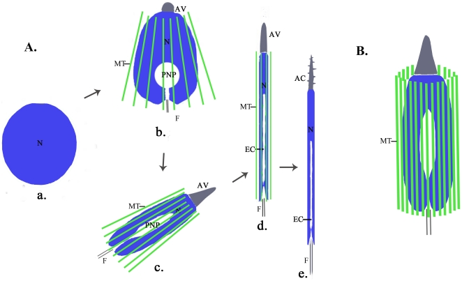
Background: Nuclear morphogenesis is one of the most fundamental cellular transformations taking place during spermatogenesis. In rodents, a microtubule-based perinuclear structure, the manchette, and a C-terminal kinesin motor KIFC1 are believed to play crucial roles in this process. Spermatogenesis in Octopus tankahkeei is a good model system to explore whether evolution has created a cephalopod prototype of mammalian manchette-based and KIFC1-dependent sperm nuclear shaping machinery.

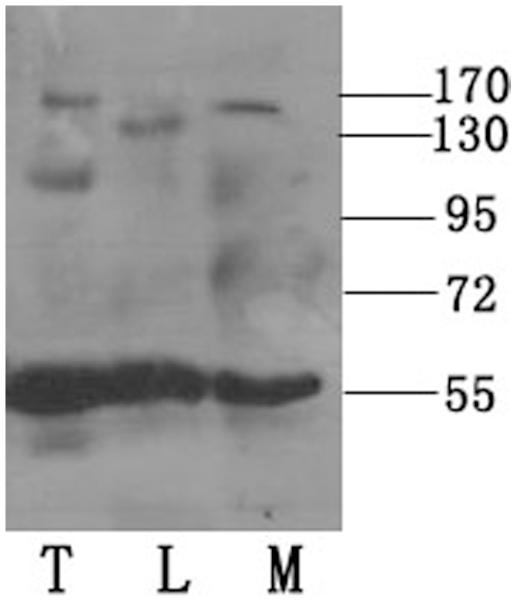
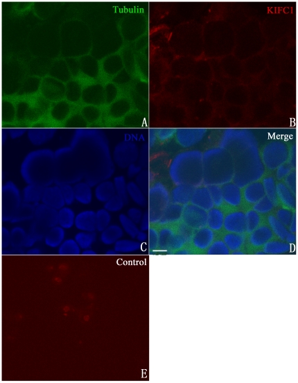
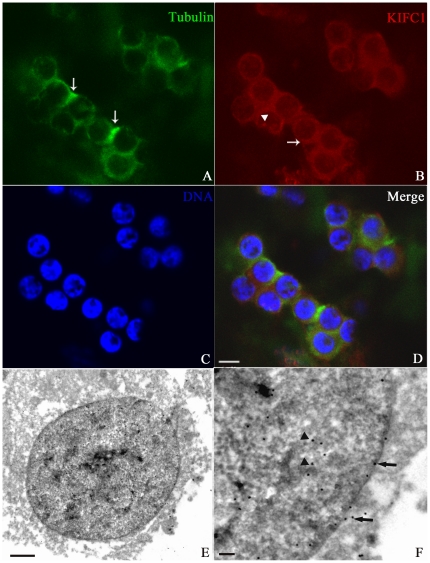
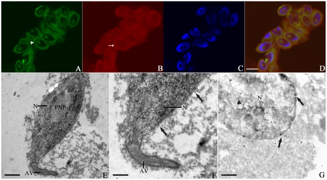
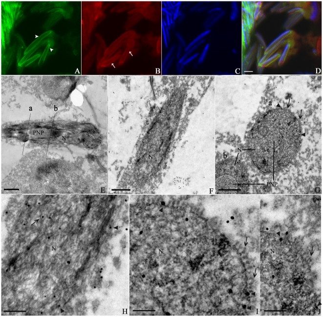
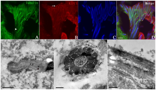
Methodology/principal findings: We detected the presence of a KIFC1-like protein in the testis, muscle, and liver of O. tankahkeei by Western Blot. Then we tracked its dynamic localization in spermatic cells at various stages using Immunofluorescence and Immunogold Electron Microscopy. The KIFC1-like protein was not expressed at early stages of spermatogenesis when no significant morphological changes occur, began to be present in early spermatid, localized around and in the nucleus of intermediate and late spermatids where the nucleus was dramatically elongated and compressed, and concentrated at one end of final spermatid. Furthermore, distribution of the motor protein during nuclear elongation and condensation overlapped with that of the cephalopod counterpart of manchette at a significant level.

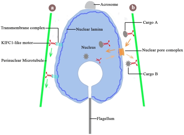
Conclusions/significance: The results support the assumption that the protein is actively involved in sperm nuclear morphogenesis in O. tankahkeei possibly through bridging the manchette-like perinuclear microtubules to the nucleus and assisting in the nucleocytoplasmic trafficking of specific cargoes. This study represents the first description of the role of a motor protein in sperm nuclear shaping in cephalopod.


Conflict of interest statement

Competing Interests: The authors have declared that no competing interests exist.

Figures

Figure 1
Figure 1. Diagrammatic representation of spermiogenesis in O. tankahkeei (diagram not to scale).
(A) Morphological differentiation of the nucleus, acrosome, and flagellum is emphasized. In early spermatid (a), the nucleus is nearly round and other major organelles do not begin to differentiate; In intermediate spermatid (b), perinuclear manchette appears, the nucleus is slightly elongated and compacted and invaginated by a posterior nuclear pocket, a coalesced acrosomal vesicle is seen at the apical-most part, and the flagellar axoneme begins to assemble; In late spermatid (c), perinuclear manchette continues to closely wrap the nucleus, the nucleus is undergoing dramatic elongation and condensation and resembles a long spindle, the acrosomal vesicle grows in size and tapers forward, and the flagellum extends in length (not shown); The final spermatid (d) is almost identical to mature spermatozoon, perinuclear manchette begins to disassemble, the nucleus adopts a long cylindrical shape with endonuclear channel formed at its posterior end, the acrosomal vesicle becomes an extended cone-like structure and will undergo a final process of helicoidization, and the flagellum is very long (not shown); The mature spermatozoon (e) has a rod-like nucleus, a screw-shaped acrosome, and a slender flagellum. Perinuclear manchette completely disappears. N: nucleus; AV: acrosomal vesicle; AC: acrosome; F: flagellum; PNP: posterior nuclear pocket; EC: endonuclear channel; MT: manchette microtubule. (B) A perinuclear microtubules-based structure analogous to mammalian manchette is formed during sperm nuclear morphogenesis in O. tankahkeei. The microtubules are considered to run parallel to the long axis of the nucleus and wrap the nucleus. MT: manchette microtubule.
Figure 2
Figure 2. Detection of the KIFC1-like protein by Western Blot.
The KIFC1-like protein was present in the testis (T), muscle (M) and liver (L) of O. tankahkeei. The protein migrated to a position on the membrane corresponding to a molecular weight of slightly above 55 kDa. No obvious difference of protein expression level was discerned from the result. Molecular weight marker (kDa) was labeled on the right.
Figure 3
Figure 3. Absence of the KIFC1-like protein at early stages of spermatogenesis in O. tankahkeei.
The protein was immuno-detected with KIFC1 antibody and Texas Red conjugated secondary antibody. Tubulin was localized using FITC-conjugated anti-tubulin antibody. DAPI was used to stain DNA. (A) Microtubules (green signal) were randomly scattered in the cytoplasm. (B) The KIFC1-like protein (red signal) was not expressed at the moment. (C) Nuclei staining (blue signal) of spermatogonia or spermatocytes. (D) A merge of the above three pictures. (E) Omission of antibody to KIFC1 was set as a negative control. Scale bar equals 5 µm.
Figure 4
Figure 4. Distribution of the KIFC1-like protein and microtubules in early spermatid in O. tankahkeei.
The KIFC1-like protein and microtubules are visualized by IF and IEM. (A) Microtubules seemed preferentially arranged near the basal zone of the spermatid head (arrow) where the manchette-like structure began to assemble. The green signal displayed varying intensity at different places and was not continuous around the nucleus. (B) Elevated expression of the KIFC1-like protein was initiated. The signal was apparent in vicinity to the nuclear envelope (arrow) and also present in the nucleus with lower intensity (arrowhead). (C) Nuclei staining of early spermatid. (D) A merge of the above three pictures. (E, F) The gold particles were located near the nuclear membranes (arrows) and in the nucleus (arrowheads). F is a higher magnification of an area in E. Scale bar is 5 µm in (A–D), 1 µm in (E) and 0.25µm in (F).
Figure 5
Figure 5. Localization of the KIFC1-like protein and microtubules in intermediate spermatid in O. tankahkeei.
The distribution of KIFC1-like protein and microtubules are observed by IF and IEM. (A) Microtubules were tightly attached to the nuclear periphery (arrowhead). (B) The KIFC1-like protein was abundantly expressed and mainly located around and in the nucleus (arrow). (C) Nuclei staining of intermediate spermatid. (D) A merge of the above three pictures. (E, F) Longitudinal sections of the spermatid. Some of the gold particles were located in proximity to the nuclear perimeter (arrows) corresponding to the growing manchette-like structure. F is a higher magnification of an area in E. (G) Transverse section of the spermatid. The gold particles were clearly seen near the nuclear envelope (arrows) as well as in the nucleus (arrowhead). Scale bar is 5 µm in (A–D), 1 µm in (E) and 0.5 µm in (F and G). AV: acrosomal vesicle. N: nucleus. PNP: posterior nuclear pocket.
Figure 6
Figure 6. Localization of the KIFC1-like protein and microtubules in late spermatid in O. tankahkeei.
The IF and IEM methods were hired to detect the expression and localization of KIFC1-like protein and microtubules at the late spermatid stage. (A) The manchette-like structure was seen encircling the nucleus (arrowheads). (B) The KIFC1-like protein was still mainly located around and in the nucleus, but the expression level was relatively decreased as the signal intensity declined (arrows). (C) Nuclei staining of late spermatid. (D) A merge of the above three pictures. (E, F, H) Longitudinal sections of a segment of the spermatid. The gold particles were located in proximity to the nuclear periphery (arrowheads) as well as in the nucleus (arrow). Cross sections at two positions labeled as “a” and “b” were presented in G. H is a higher magnification of an area in F. (G, I, J) Transverse sections of the spermatid. Some gold particles (arrowheads) were distributed between the nuclear envelope and the manchette-like perinuclear structure (arrows) which exhibited a transverse profile of a circle of holes closely attached to the nuclear periphery. I and J are higher magnifications of an area of “a” and “b” labled in G, respectively. Scale bar is 5 µm in (A–D), 1 µm in (E-G), 0.5 µm in (H) and 0.25 µm in (I and J). N: nucleus. PNP: posterior nuclear pocket.
Figure 7
Figure 7. Localization of the KIFC1-like protein and microtubules in final spermatid in O. tankahkeei.
The location of KIFC1-like protein and microtubules were checked by IF and IEM. (A) Microtubules were still visible in the cell but the manchette-like perinuclear structure was not so rigid around the nucleus (arrowhead). (B) Signal intensity of the KIFC1-like protein greatly decreased, indicating its expression level significantly dropped. The signal around and in the nucleus was not as apparent as seen in previous stages. Most of the protein would concentrate like a dot at one end (arrow) of the spermatid probably corresponding to the caudal region where residual cytoplasm was disposed. (C) Nuclei staining of final spermatid. (D) A merge of the above three pictures. (E) Longitudinal section of the maturing acrosome. The gold particles were present in the inner striated structure (arrowheads) and near the helical surface (arrows). (F) Transverse section of the spermatid. The manchette-like perinuclear microtubules were absent. The gold particles were seen near the nuclear envelope (arrows) and in the nucleus (arrowhead) with low density. (G) Longitudinal section of the spermatid. The gold particles were sparse in the cell, implying significant decline of protein expression. Scale bar is 5 µm in (A–D), 1 µm in (E and G), and 0.25 µm in (F). AV: acrosomal vesicle. N: nucleus. EC: endonuclear channel.
Figure 8
Figure 8. Two possible functional models of the KIFC1-like protein in sperm nuclear morphogenesis in O. tankahkeei.
(a) The manchette-like perinuclear microtubules are connected to the nuclear lamina inside the nucleus through association of the KIFC1-like protein with some multi-molecular complex (transmembrane complex) scanning the nuclear membranes and reaching the nuclear lamina. The KIFC1-like protein moving towards the caudal end of spermatid will transmit the motor-generated force to the nucleus and facilitate mechanical rearrangement of the nuclear lamina and other endonuclear components and eventually elongation and condensation of the nucleus. (b) The KIFC1-like protein is able to carry some structural and functional elements in vicinity to the nuclear perimeter and shuttle across the nuclear pore complex to enter or exit the nucleus via association with the nucleocytoplasmic trafficking machinery. As a result, it will assist in the import (cargo A) and export (cargo B) of a specific group of cargoes probably involved in the regulation of nuclear activities such as chromatin condensation.

Similar articles

Cited by

References

    1. Hess RA, Renato FL. Spermatogenesis and cycle of the seminiferous epithelium. Adv Exp Med Biol. 2008;636:1–15. - PubMed
    1. Hermo L, Pelletier RM, Cyr DG, Smith CE. Surfing the wave, cycle, life history, and genes/proteins expressed by testicular germ cells. Part 1: background to spermatogenesis, spermatogonia, and spermatocytes. Microsc Res Tech. 2010a;73:241–278. - PubMed
    1. Gimenez-Bonafé P, Ribes E, Zamora MJ, Kasinsky HE, Chiva M. Evolution of octopod sperm I: comparison of nuclear morphogenesis in Eledone and Octopus. Mol Reprod Dev. 2002;62:357–362. - PubMed
    1. Hermo L, Pelletier RM, Cyr DG, Smith CE. Surfing the wave, cycle, life history, and genes/proteins expressed by testicular germ cells. Part 2: changes in spermatid organelles associated with development of spermatozoa. Microsc Res Tech. 2010b;73:279–319. - PubMed
    1. Dadoune JP, Siffroi JP, Alfonsi MF. Transcription in haploid male germ cells. Int Rev Cytol. 2004;237:1–56. - PubMed

Publication types