. 2010:2010:395758.
doi: 10.1155/2010/395758. Epub 2010 Apr 6.

The Cell Wall Teichuronic Acid Synthetase (TUAS) Is an Enzyme Complex Located in the Cytoplasmic Membrane of Micrococcus luteus

Affiliations

The Cell Wall Teichuronic Acid Synthetase (TUAS) Is an Enzyme Complex Located in the Cytoplasmic Membrane of Micrococcus luteus

Lingyi Lynn Deng et al. Biochem Res Int. 2010.

Abstract

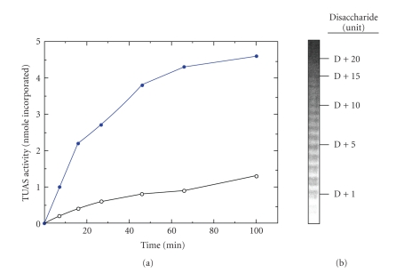
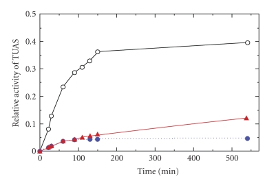
The cell wall teichuronic acid (TUA) of Micrococcus luteus is a long-chain polysaccharide composed of disaccharide repeating units [-4-β-D-ManNAcAp-(1→6)α-D-Glcp-1-](n), which is covalently anchored to the peptidoglycan on the inner cell wall and extended to the outer surface of the cell envelope. An enzyme complex responsible for the TUA chain biosynthesis was purified and characterized. The 440 kDa enzyme complex, named teichuronic acid synthetase (TUAS), is an octomer composed of two kinds of glycosyltransferases, Glucosyltransferase, and ManNAcA-transferase, which is capable of catalyzing the transfer of disaccharide glycosyl residues containing both glucose and the N-acetylmannosaminuronic acid residues. TUAS displays hydrophobic properties and is found primarily associated with the cytoplasmic membrane. The purified TUAS contains carotinoids and lipids. TUAS activity is diminished by phospholipase digestion. We propose that TUAS serves as a multitasking polysaccharide assembling station on the bacterial membrane.


Figures

Figure 1
Figure 1
Chemical structure of teichuronic acid in the cell wall of Micrococcus luteus and proposed site of action for glucosyltransferase (indicated by arrow 1) and that for ManNAcA transferase (indicated by arrow 2). The linker contains an oligosaccharide with a GlcNAc residue at the reducing end and a phosphate.
Figure 2
Figure 2
(a) Effect of the two substrates (UDP-[14C]glucose and UDP-ManNAcA) on the activity of teichuronic acid synthetase (TUAS). Open circle: with one substrate 0.5 mM UDP-C14-Glucose; Closed circle: two substrates by adding 0.5 mM UDP-ManNAcA to the same mix. (b) Analysis of teichuronic acid, the enzymatic product of TUAS, by nondenaturing PAGE stained with Alcian blue and silver. The numbers on the right represent the repeats of the basic TUA disaccharide unit. D is an oligosaccharide of TUA estimated to have 6–10 disaccharide repeats, which is not detectable by PAGE.
Figure 3
Figure 3
(a) M. luteus TUAS analyzed with native gradient PAGE. Partially purified TUAS was subjected to electrophoresis on 3–12% native gradient polyacrylamide gels (left, lane 2). Protein bands were visualized by staining with Coomassie blue. The molecular-mass standards of 545, 272, 132, and 66 kDa (top to bottom) are shown (left, lane 1). The TUAS activity is indicated with an arrow. (b) SDS-PAGE of M. luteus TUAS. Protein bands separated by 9.5% SDS-PAGE were visualized by silver staining. Lane 1: Molecular-mass standards of 97.4, 66.2, 42.7, and 31 kDa, respectively (top to bottom). Lane 2: Detergent extraction of membrane fraction. Lane 3: Protein after DEAE-cellulose column chromatography. Lane 4: TUAS purified to near homogeneity by isoelectric precipitation (pH 5.0) after DEAE-cellulose, gel filtration and adsorbent column chromatography. TUAS has two subunits A and B, upper subunit (A) is equivalent to 54 kDa and the lower subunit (B) to 52.5 kDa.
Figure 4
Figure 4
Time course of TUAS activity of M. luteus and the effect of phospholipase C on the activity. Open circle: TUAS activity; Closed circle: TUAS treated with phospholipase C; Triangle: addition of artificial phospholipid vesicles to TUAS reaction mix following phospholipase treatment.
Figure 5
Figure 5
Immunoflluorescent images of M. Luteus. For (a) and (b), cells were observed under a fluorescent microscope with excitation wavelength 495 nm and emission wavelength 520 nm. (a) a negative control in which M. luteus cells were incubated with the secondary antibody conjugated to FITC. (b) M. luteus cells were incubated with both anti-TUAS serum and the secondary antibody conjugated to FITC. (c) The same bacterial cells as (b) except under visible light.
Figure 6
Figure 6
Proposed mode of action and subcellular location for cell wall TUA biosynthesis, in which an enzyme complex, TUAS, consisting of two kinds of enzymes (Glucosyltransferase and ManNAcA-transferase), serves as a multitasking polysaccharide assembling station on the bacterial membrane for efficient elongation of cell wall teichuronic acid. Black Diamond: ManNAcA, White Circle: glucose, Rectangle: linkage oligosaccharide, and Yellow Circle: phosphate.

References

    1. Hase S, Matsushima Y. Structural studies on a glucose-containing polysaccharideobtained from Micrococcus lysodeikticus cell walls. IV. Isolation of a disaccharide, 6-O-(strontium 2-acetamido-2-deoxy-beta-D-mannopyranosyluronate)-D-glucose. The Journal of Biochemistry. 1972;72(5):1117–1128. - PubMed
    1. Johnson SD, Lacher KP, Anderson JS. Carbon-13 nuclear magnetic resonance spectroscopic study of teichuronic acid from Micrococcus luteus cell walls. Comparison of the polysaccharide isolated from cells with that synthesized in vitro. Biochemistry. 1981;20(16):4781–4785. - PubMed
    1. Johnson GL, Hoger JH, Ratnayake JH, Anderson JS. Characterization of three intermediates in the biosynthesis of teichuronic acid of Micrococcus luteus . Archives of Biochemistry and Biophysics. 1984;235(2):679–691. - PubMed
    1. Gassner GT, Dickie JP, Damerski DA, Magnuson JK, Anderson JS. Teichuronic acid reducing terminal N-acetylglucosamine residue linked by phosphodiester to peptidoglycan of Micrococcus luteus . Journal of Bacteriology. 1990;172(5):2273–2279. - PMC - PubMed
    1. Traxler CI, Goustin AS, Anderson JS. Elongation of teichuronic acid chains by a wall-membrane preparation from Micrococcus luteus . Journal of Bacteriology. 1982;150(2):649–656. - PMC - PubMed

LinkOut - more resources