

Primary retroperitoneal paraganglioma simulating a pancreatic mass: a case report and review of the literature
- PMID: 21188160
- PMCID: PMC3004405
- DOI: 10.1155/2010/645728
Primary retroperitoneal paraganglioma simulating a pancreatic mass: a case report and review of the literature
Abstract
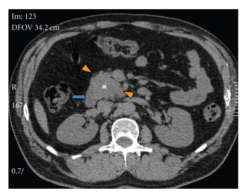
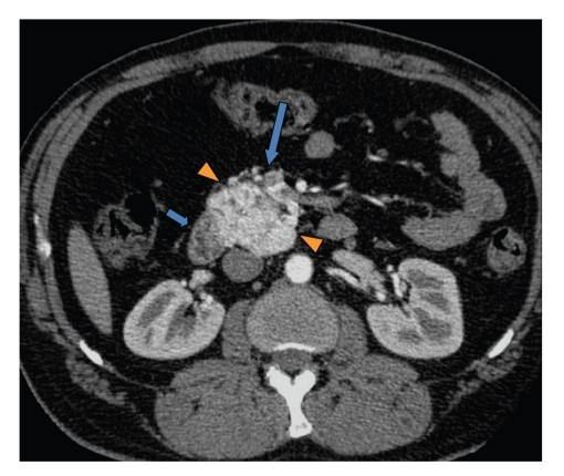
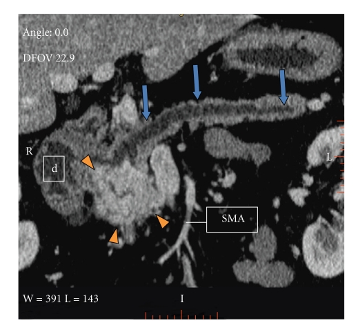
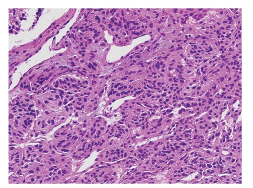
Paragangliomas are extra-adrenal tumors of the autonomic nervous system and may be found within the skull base, neck, chest, and abdomen. When presenting within the abdominal cavity, they may arise as a primary retroperitoneal neoplasm and can mimic vascular malformations or other conditions related to specific retroperitoneal organs such as the pancreas, kidneys, or adrenals. Retroperitoneal paragangliomas are mostly benign with good prognosis; however, they can present with abdominal pain, palpable mass, or hypertensive episodes. Patients should be initially evaluated with catecholamine levels, followed by computed tomography or magnetic resonance imaging to locate the primary lesion. Surgical excision remains the mainstay of treatment, although advanced disease and proximity to vital organs can make excision difficult or impossible. This case report describes a patient who initially underwent work up for a suspected pancreatic head mass which was discovered to be a retroperitoneal paraganglioma by frozen section.
Figures
References
-
- Nishino M, Hayakawa K, Minami M, Yamamoto A, Ueda H, Takasu K. Primary retroperitoneal neoplasms: CT and MRI imaging findings with anatomic and pathologic diagnostic clues. Radiographics. 2003;23(1):45–57. - PubMed
-
- Rha SE, Byun JY, Jung SE, Chun HJ, Lee HG, Lee JM. Neurogenic tumors in the abdomen: tumor types and imaging characteristics. Radiographics. 2003;23(1):29–43. - PubMed
-
- Yang JH, Bae SJ, Park S, et al. Bilateral pheochromocytoma associated with paraganglioma and papillary thyroid carcinoma: report of an unusual case. Endocrine Journal. 2007;54(2):227–231. - PubMed
-
- Kaltsas GA, Besser GM, Grossman AB. The diagnosis and medical management of advanced neuroendocrine tumors. Endocrine Reviews. 2004;25(3):458–511. - PubMed
-
- Elder EE, Elder G, Larsson C. Pheochromocytoma and functional paraganglioma syndrome: no longer the 10% tumor. Journal of Surgical Oncology. 2005;89(3):193–201. - PubMed
Publication types
MeSH terms
LinkOut - more resources
Full Text Sources
Medical
