

Peyer's Patches: The Immune Sensors of the Intestine
- PMID: 21188221
- PMCID: PMC3004000
- DOI: 10.4061/2010/823710
Peyer's Patches: The Immune Sensors of the Intestine
Abstract
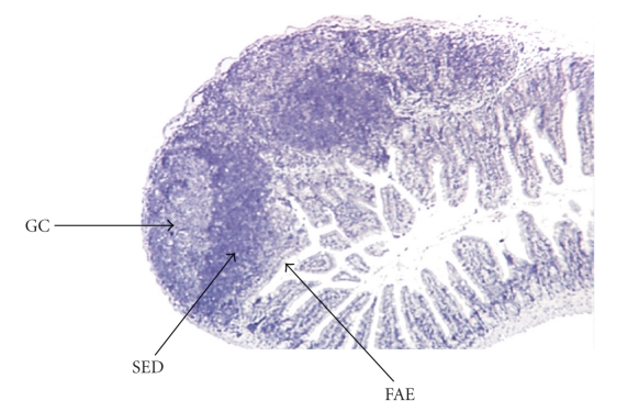
The gut-associated lymphoid tissue (GALT) consists of isolated or aggregated lymphoid follicles forming Peyer's patches (PPs). By their ability to transport luminal antigens and bacteria, PPs can be considered as the immune sensors of the intestine. PPs functions like induction of immune tolerance or defense against pathogens result from the complex interplay between immune cells located in the lymphoid follicles and the follicle-associated epithelium. This crosstalk seems to be regulated by pathogen recognition receptors, especially Nod2. Although TLR exerts a limited role in PP homeotasis, Nod2 regulates the number, size, and T-cell composition of PPs, in response to the gut flora. In turn, CD4(+) T-cells present in the PP are able to modulate the paracellular and transcellular permeabilities. Two human disorders, Crohn's disease and graft-versus-host disease are thought to be driven by an abnormal response toward the commensal flora. They have been associated with NOD2 mutations and PP dysfunction.
Figures
References
-
- Neutra MR, Mantis NJ, Kraehenbuhl J-P. Collaboration of epithelial cells with organized mucosal lymphoid tissues. Nature Immunology. 2001;2(11):1004–1009. - PubMed
-
- Van Kruiningen HJ, Ganley LM, Freda BJ. The role of Peyer’s patches in the age-related incidence of Crohn’s disease. Journal of Clinical Gastroenterology. 1997;25(2):470–475. - PubMed
-
- Van Kruiningen HJ, West AB, Freda BJ, Holmes KA. Distribution of Peyer’s patches in the distal ileum. Inflammatory Bowel Diseases. 2002;8(3):180–185. - PubMed
-
- Kyriazis AA, Esterly JR. Fetal and neonatal development of lymphoid tissues. Archives of Pathology. 1971;91(5):444–451. - PubMed
LinkOut - more resources
Full Text Sources
Other Literature Sources
Research Materials
Miscellaneous
