. 2010:2010:751313.
doi: 10.1155/2010/751313. Epub 2010 Dec 5.

Ivabradine reduces chemokine-induced CD4-positive lymphocyte migration

Affiliations

Ivabradine reduces chemokine-induced CD4-positive lymphocyte migration

Thomas Walcher et al. Mediators Inflamm. 2010.

Abstract

Aims: Migration of CD4-positive lymphocytes into the vessel wall is a critical step in atherogenesis. Recent data suggest that ivabradine, a selective I(f)-channel blocker, reduces atherosclerotic plaque formation in apolipoprotein E-deficient mice, hitherto nothing is known about the mechanism by which ivabradine modulates plaque formation. Therefore, the present study investigated whether ivabradine regulates chemokine-induced migration of lymphocytes.

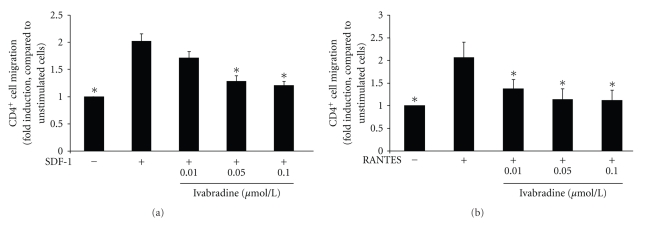
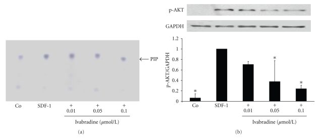
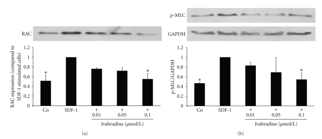
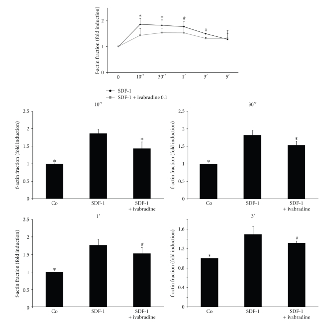
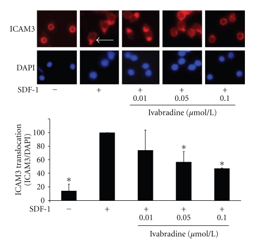
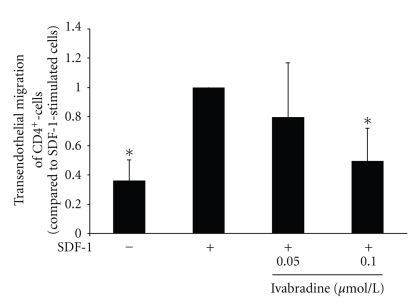
Methods and results: Stimulation of CD4-positive lymphocytes with SDF-1 leads to a 2.0 ± 0.1 fold increase in cell migration (P < .01; n = 7). Pretreatment of cells with ivabradine reduces this effect to a maximal 1.2 ± 0.1 fold induction at 0.1 µmol/L ivabradine (P < .01 compared to SDF-1-treated cells, n = 7). The effect of ivabradine on CD4-positive lymphocyte migration was mediated through an early inhibition of chemokine-induced PI-3 kinase activity as determined by PI-3 kinase activity assays. Downstream, ivabradine inhibits activation of the small GTPase Rac and phosphorylation of the Myosin Light Chain (MLC). Moreover, ivabradine treatment reduces f-actin formation as well as ICAM3 translocation to the uropod of the cell, thus interfering with two important steps in T cell migration.

Conclusion: Ivabradine inhibits chemokine-induced migration of CD4-positive lymphocytes. Given the crucial importance of chemokine-induced T-cell migration in early atherogenesis, ivabradine may be a promising tool to modulate this effect.


Figures

Figure 1
Figure 1
Ivabradine reduces SDF-1 and RANTES-induced CD4-positive lymphocyte migration. (a) Human CD4-positive cells were pretreated with ivabradine for 15 minutes at concentrations indicated before migration experiments using SDF-1 (100 ng/mL) were performed in a modified Boyden chamber. Data are expressed as fold induction compared to SDF-1-stimulated cells. Bars represent mean ± SD (n = 7); P < .01 compared to chemokine-stimulated cells. (b) Human CD4-positive lymphocytes were pretreated with ivabradine for 15 minutes at concentrations indicated before migration experiments using RANTES (100 pg/ml) were performed. Data are expressed as fold induction of chemokine-stimulated cells. Bars represent mean ± SD (n = 7); *P < .01 compared to chemokine-stimulated cells.
Figure 2
Figure 2
Ivabradine inhibits SDF-1-induced PI 3-kinase activity and phosphorylation of AKT. (a) Human CD4-positive cells were pretreated with ivabradine in different concentrations for 15 minutes before cells were stimulated with SDF-1 (100 ng/mL). After 5 minutes, PI 3-kinase activity assay was performed. Specific dots are labelled with an arrow (PIP). Three independent experiments showed similar results. (b) SDF-1 leads to phosphorylation of AKT. Isolated CD4-positive lymphocytes were pretreated with ivabradine in different concentrations indicated before stimulation with 100 ng/mL SDF-1 for 10 min. Total lysates were analyzed by immunoblotting employing antibodies against phospho-AKT. Equal loading of intact protein was confirmed by staining for GAPDH. Densitometric analysis were performed of 3 independent experiments. Data are expressed as p-AKT normalized to GAPDH. Bars represent mean ± SD. *P < .01 compared with SDF-1-stimulated cells; n = 3.
Figure 3
Figure 3
Ivabradine inhibits SDF-1-induced activation of Rac1 and reduces phosphorylation of MLC. (a) Human CD4-positive cells were pretreated with ivabradine (0.01, 0.05, and 0.1 μmol/L) for 15 minutes before cells were stimulated with SDF-1 (100 ng/mL). After 3 minutes, GTPase activity assay was performed (affinity precipitation with GST-PAK). Densitometric analysis of 5 independent experiments. Bars represent mean ± SD. *P < .01 compared with SDF-1-stimulated cells. (b) SDF-1 leads to phosphorylation of MLC. Isolated CD4-positive lymphocytes were pretreated with ivabradine in different concentrations indicated before stimulation with 100 ng/mL SDF-1 for 5 min. Total lysates were analyzed by immunoblotting employing antibodies against phospho-MLC. Equal loading of intact protein was confirmed by staining for GAPDH. Densitometric analyses were performed of 5 independent experiments. Data are expressed as p-MLC normalized to GAPDH. Bars represent mean ± SD. *P < .01 compared with SDF-1-stimulated cells; n = 5.
Figure 4
Figure 4
Ivabradine reduces actin polymerisation in CD4-positive lymphocytes. (a) Isolated lymphocytes were pretreated with ivabradine (n = 5) for 15 min before stimulation with SDF-1. Actin polymerisation was determined by flow cytometry at times indicated. Lower panel represents statistical analysis for the four timepoints indicated. Bars represent mean ± SD; n = 5; # P < .05; *P < .01 compared to SDF-1-stimulated cells.
Figure 5
Figure 5
Ivabradine abolishes SDF-1-induced ICAM3 translocation. CD4-positive lymphocytes were pretreated with ivabradine (0.01, 0.05, or 0.1 μmol/L) before stimulation with SDF-1 for 30 min. ICAM3 translocation was assayed using immunofluorescence staining. ICAM3 translocation at the uropod of migrating cells is indicated by the arrow. Lower panel shows statistical analysis of cells positive for ICAM3 translocation as % of DAPI-positive cells; bars represent mean ± SD; n = 6; *P < .01 compared to SDF-1-stimulated cells.
Figure 6
Figure 6
Ivabradine reduces SDF-1-induced transendothelial migration of CD4-positive lymphocytes. Human CD4-positive cells were pretreated with ivabradine for 15 minutes at 0.05 or 0.1 μmol/L before migration experiments using SDF-1 (100 ng/mL) were performed in a transendothelial migration assay. Data are expressed as fold induction compared to SDF-1-stimulated cells. Bars represent mean ± SD (n = 6); P < .01 compared to chemokine-stimulated cells.

References

    1. Hansson GK. Mechanisms of disease: inflammation, atherosclerosis, and coronary artery disease. New England Journal of Medicine. 2005;352(16):1685–1626. - PubMed
    1. Zernecke A, Weber C. Inflammatory mediators in atherosclerotic vascular disease. Basic Research in Cardiology. 2005;100(2):93–101. - PubMed
    1. Heller EA, Liu E, Tager AM, et al. Chemokine CXCL10 promotes atherogenesis by modulating the local balance of effector and regulatory T cells. Circulation. 2006;113(19):2301–2312. - PubMed
    1. Veillard NR, Steffens S, Pelli G, et al. Differential influence of chemokine receptors CCR2 and CXCR3 in development of atherosclerosis in vivo. Circulation. 2005;112(6):870–878. - PubMed
    1. Kannel WB, Kannel C, Paffenbarger RS, Jr., Cupples A. Heart rate and cardiovascular mortality: the Framingham study. American Heart Journal. 1987;113(6):1489–1494. - PubMed

Publication types

MeSH terms