Review
. 2010 Dec;11(4):219-27.
doi: 10.1007/s11154-010-9153-1.

Cellular mechanisms of bone remodeling

Affiliations
Review

Cellular mechanisms of bone remodeling

Erik Fink Eriksen. Rev Endocr Metab Disord. 2010 Dec.

Abstract

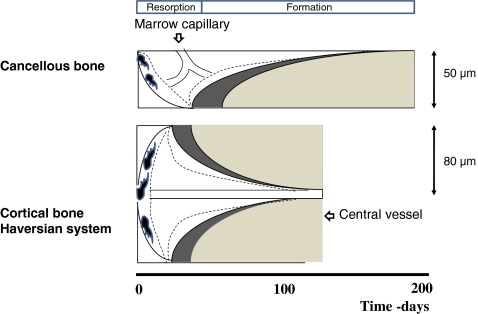
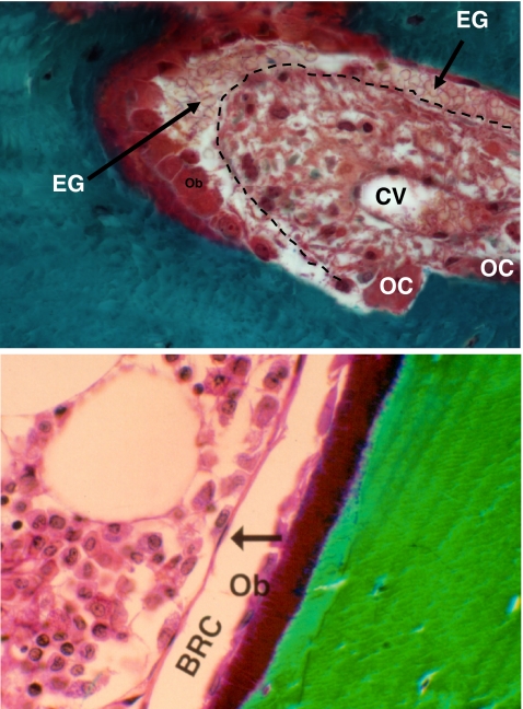
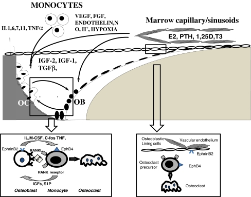
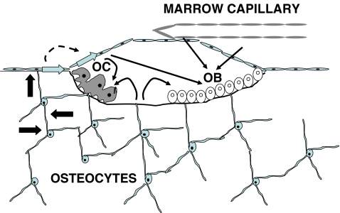
Bone remodeling is a tightly regulated process securing repair of microdamage (targeted remodeling) and replacement of old bone with new bone through sequential osteoclastic resorption and osteoblastic bone formation. The rate of remodeling is regulated by a wide variety of calcitropic hormones (PTH, thyroid hormone, sex steroids etc.). In recent years we have come to appreciate that bone remodeling proceeds in a specialized vascular structure,--the Bone Remodeling Compartment (BRC). The outer lining of this compartment is made up of flattened cells, displaying all the characteristics of lining cells in bone including expression of OPG and RANKL. Reduced bone turnover leads to a decrease in the number of BRCs, while increased turnover causes an increase in the number of BRCs. The secretion of regulatory factors inside a confined space separated from the bone marrow would facilitate local regulation of the remodeling process without interference from growth factors secreted by blood cells in the marrow space. The BRC also creates an environment where cells inside the structure are exposed to denuded bone, which may enable direct cellular interactions with integrins and other matrix factors known to regulate osteoclast/osteoblast activity. However, the denuded bone surface inside the BRC also constitutes an ideal environment for the seeding of bone metastases, known to have high affinity for bone matrix. Circulating osteoclast- and osteoblast precursor cells have been demonstrated in peripheral blood. The dominant pathway regulating osteoclast recruitment is the RANKL/OPG system, while many different factors (RUNX, Osterix) are involved in osteoblast differentiation. Both pathways are modulated by calcitropic hormones.


Figures

Fig. 1
Fig. 1
Schematic representation of Bone Multicellular Units (BMUs) in cancellous and cortical bone. Broken lines denote the outer limit of Bone Remodeling Compartment associated with the resorptive and formative sites of the BMU. The mean thickness of the structure in cancellous bone is 50 μm and 80 μm in cortical bone equivalent to a mean Haversian system diameter of 160 μm. The Blood supply for the BRCs is provided by capillaries either coming from the marrow space as is the case for cancellous BMUs or from the central vessel of Haversian systems in cortical bone. The duration of the remodeling sequence is somewhat longer in cancellous than in cortical bone. The position of marrow cappillaries is hypothetical, as the exact distribution is poorly elucidated
Fig. 2
Fig. 2
Different representations of BRC structures in cortical (upper panel) and trabecular bone (lower panel). In cortical bone the BRC (outer demarcation by the broken line) is filled with erythrocyte ghosts (EG) and is located at the closing cone of the Haversian system situated over osteoblasts (OB). A few osteoclasts (OC) are also seen. CV denotes the central vessel of the Haversian system. In trabecular bone (lower panel) the outer lining of the BRC is clearly discernible, demarcating a vascular structure on top of osteoblasts (OB). Picture in upper panel courtesy of Pierre Delmas, Lyon, France
Fig. 3
Fig. 3
Depiction of some of the main local regulatory factors operating at remodeling sites with osteoclasts (OC) and osteoblasts (OB). Interleukins (IL), tumor necrosis factors (TNF), transforming growth factors (TGF), colony stimulating factors (CSF), Insulin like growth factors (IGF), fibroblast growth factors (FGF), platelet derived growth factors (PDGF), bone morphogenetic proteins (BMP)) are formed by both monocytic cells in the marrow space or circulation, as well as bone cells in the BMU. NFκB- or RANK- ligand (RANKL) and osteoprotegerin (OPG) are formed specifically by osteoblasts. Factors from the marrow space as well as factors liberated by endothelial cells (vascular endothelial growth factor (VEGF), endothelin, nitrogen oxide (NO)) may diffuse to receptors on osteoclasts or osteoblasts. The cellular responses in the BMU are then further modulated by systemic hormones in the circulation (estrogen (E2), parathyroid hormone (PTH), active vitamin D (1,25D), thyroid hormone (T3)). Left lower insert depicts in detail osteoblast-osteoclast interactions inside the BRC and right lower insert depict an alternativ, still hypohetical, version of that interaction based on lining cells acting as the osteoblastic component in thata interaction
Fig. 4
Fig. 4
Connections between the osteocyte network, lining cells and the BRC. All cells in this network are connected with gap junctions, which may provide a pathway (block arrows), by which signals generated deep within bone may reach the surface and elicit remodeling events by osteoclasts (OC) and osteoblasts (OB) in response to mechanical stimuli. The response may be modulated by factors liberated from the vascular endothelium or marrow capillaries/sinusoids and paracrine factors (broken arrow) liberated from lining cells may also play a role

References

    1. Frost HM. Tetracycline-based histological analysis of bone remodeling. Calcif Tissue Res. 1969;3:211–237. doi: 10.1007/BF02058664. - DOI - PubMed
    1. Eriksen EF. Normal and pathological remodeling of human trabecular bone: three dimensional reconstruction of the remodeling sequence in normals and in metabolic bone disease. Endocr Rev. 1986;7:379–408. doi: 10.1210/edrv-7-4-379. - DOI - PubMed
    1. Eriksen EF, Gundersen HJ, Melsen F, Mosekilde L. Reconstruction of the formative site in iliac trabecular bone in 20 normal individuals employing a kinetic model for matrix and mineral apposition. Metab Bone Dis Relat Res. 1984;5:243–252. doi: 10.1016/0221-8747(84)90066-3. - DOI - PubMed
    1. Eriksen EF, Melsen F, Mosekilde L. Reconstruction of the resorptive site in iliac trabecular bone: a kinetic model for bone resorption in 20 normal individuals. Metab Bone Dis Relat Res. 1984;5:235–242. doi: 10.1016/0221-8747(84)90065-1. - DOI - PubMed
    1. Eriksen EF, Hodgson SF, Eastell R, Cedel SL, O’Fallon WM, Riggs BL. Cancellous bone remodeling in type I (postmenopausal) osteoporosis: quantitative assessment of rates of formation, resorption, and bone loss at tissue and cellular levels. J Bone Miner Res. 1990;5:311–319. doi: 10.1002/jbmr.5650050402. - DOI - PubMed