. 2011 Jun;7(2):471-7.
doi: 10.1007/s12015-010-9224-4.

An improved technique for chromosomal analysis of human ES and iPS cells

Affiliations

An improved technique for chromosomal analysis of human ES and iPS cells

Daniela Moralli et al. Stem Cell Rev Rep. 2011 Jun.

Abstract

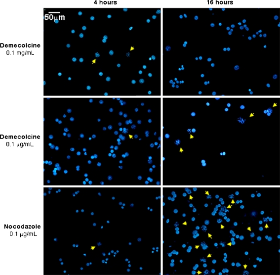
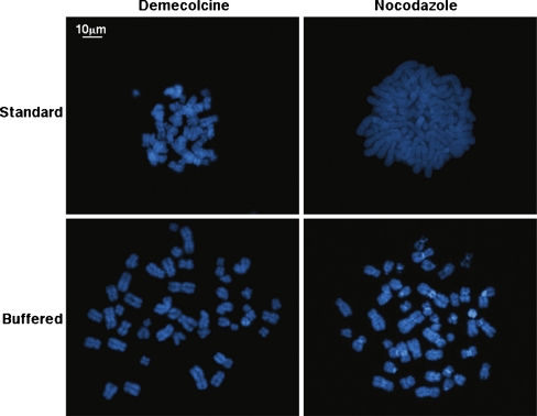
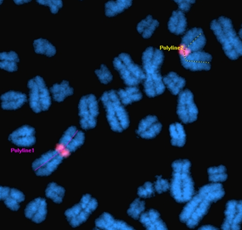
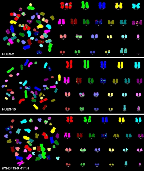
Prolonged in vitro culture of human embryonic stem (hES) cells can result in chromosomal abnormalities believed to confer a selective advantage. This potential occurrence has crucial implications for the appropriate use of hES cells for research and therapeutic purposes. In view of this, time-point karyotypic evaluation to assess genetic stability is recommended as a necessary control test to be carried out during extensive 'passaging'. Standard techniques currently used for the cytogenetic assessment of ES cells include G-banding and/or Fluorescence in situ Hybridization (FISH)-based protocols for karyotype analysis, including M-FISH and SKY. Critical for both banding and FISH techniques are the number and quality of metaphase spreads available for analysis at the microscope. Protocols for chromosome preparation from hES and human induced pluripotent stem (hiPS) cells published so far appear to differ considerably from one laboratory to another. Here we present an optimized technique, in which both the number and the quality of chromosome metaphase spreads were substantially improved when compared to current standard techniques for chromosome preparations. We believe our protocol represents a significant advancement in this line of work, and has the required attributes of simplicity and consistency to be widely accepted as a reference method for high quality, fast chromosomal analysis of human ES and iPS cells.


Figures

Fig. 1
Fig. 1
Efficiency of mitotic arrest following treatment with either demecolcine or nocodazole. Different concentrations and incubation times were compared. After fixation the cells were stained with DAPI, and analyzed at the microscope. Ten random fields from each of the slides prepared under different conditions were captured with Genus on the CytoVision system. The yellow arrows identify metaphasic cells
Fig. 2
Fig. 2
Assessment of chromosome overlaps following different mitotic and hypotonic treatments. The number of chromosome overlaps per metaphase was compared in four different ‘harvest’ conditions. Where the metaphase quality was poor, we assigned an arbitrary number of 23 overlaps per metaphase, meaning that all the chromosomes were involved in at least one overlapping event
Fig. 3
Fig. 3
Chromosome length as a parameter for metaphase spread quality for karyotype analysis. The average length of chromosome 1 (here with the centromeric region marked in red) was measured using the image analysis package MetaMorph v7.6 (example above), and compared between the nocodazole 16 h/buffered hypotonic harvest and the demecolcine 16 h/buffered hypotonic harvest
Fig. 4
Fig. 4
Twenty-four color karyotyping of hES cells (HUES-2 and HUES-10) and iPS-DF19-9-11T.H by M-FISH. The high standard and improved speed of the M-FISH analysis have together confirmed the newly identified optimal mitotic arrest and hypotonic conditions to provide a significant technical breakthrough for chromosomal analysis of hES and hiPS cells. While HUES-10 (passage 37) and iPS-DF19-9-11T.H (passage 29) presented a normal karyotype, M-FISH analysis on HUES-2 at passage 40 revealed, as well as chromosome 12 partial trisomy, a couple of structural abnormalities to include a translocation involving an extra copy of chromosome 1q and chromosome 18, and an unbalanced translocation involving chromosomes 17 and 22

References

    1. Hoffman LM, Carpenter MK. Characterization and culture of human embryonic stem cells. Nature Biotechnology. 2005;23:699–708. doi: 10.1038/nbt1102. - DOI - PubMed
    1. Baker DE, Harrison NJ, Maltby E, Smith K, Moore HD, et al. Adaptation to culture of human embryonic stem cells and oncogenesis in vivo. Nature Biotechnology. 2007;25:207–215. doi: 10.1038/nbt1285. - DOI - PubMed
    1. Meisner LF, Johnson JA. Protocols for cytogenetic studies of human embryonic stem cells. Methods. 2008;45:133–141. doi: 10.1016/j.ymeth.2008.03.005. - DOI - PubMed
    1. Caisander G, Park H, Frej K, Lindqvist J, Bergh C, et al. Chromosomal integrity maintained in five human embryonic stem cell lines after prolonged in vitro culture. Chromosome Research. 2006;14:131–137. doi: 10.1007/s10577-006-1019-8. - DOI - PubMed
    1. Grandela C, Wolvetang E. hESC adaptation, selection and stability. Stem Cell Reviews. 2007;3:183–191. doi: 10.1007/s12015-007-0008-4. - DOI - PubMed

Publication types

MeSH terms

LinkOut - more resources