

Meta-analysis of quantitative diffusion-weighted MR imaging in the differential diagnosis of breast lesions
- PMID: 21189150
- PMCID: PMC3024311
- DOI: 10.1186/1471-2407-10-693
Meta-analysis of quantitative diffusion-weighted MR imaging in the differential diagnosis of breast lesions
Abstract
Background: To determine, in a meta-analysis, the diagnostic performance of quantitative diffusion-weighted (DW) MR imaging in patients with breast lesions.
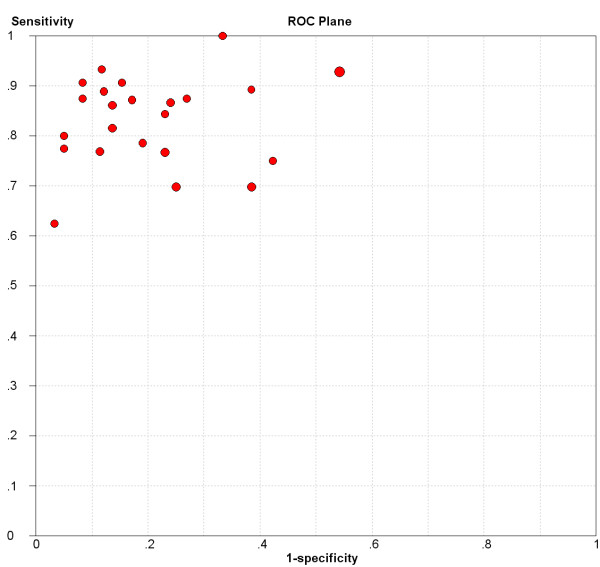
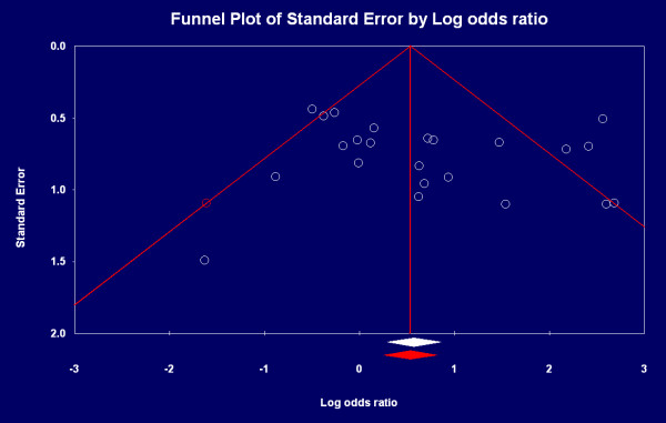
Methods: English and Chinese studies published prior to June 2009 to assess the diagnostic performance of quantitative DWI in patients with breast lesions were reviewed and summarized with reference to the inclusion and exclusion criteria. Methodological quality was assessed by using the quality assessment of diagnostic studies (QUADAS) instrument. Publication bias analysis was performed by using Comprehensive Meta-analysis version 2. Meta-Disc version 1.4 was used to describe primary results and explore homogeneity by Chi-square test and inconsistency index; to explore threshold effect by receiver operator characteristic (ROC) space and Spearman correlation coefficient; and to pool weighted sensitivity and specificity by fixed or random effect model. A summary ROC (sROC) curve was constructed to calculate the area under the curve (AUC).
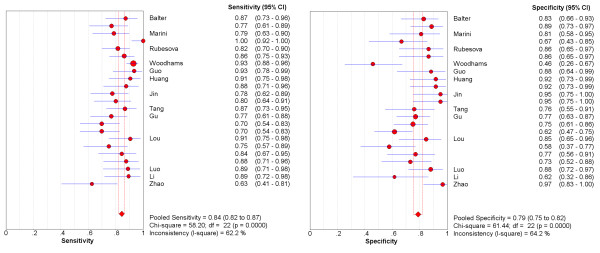
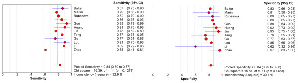
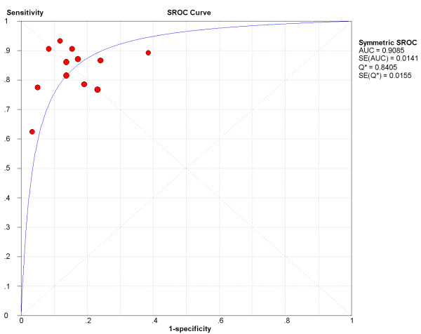
Results: Of 65 eligible studies, 13 with 615 malignant and 349 benign lesions were included in the original meta-analysis, among which heterogeneity arising from factors other than threshold effect and publication bias was explored. Methodological quality was moderate. The pooled weighted sensitivity and specificity with corresponding 95% confidence interval (CI) in one homogenous subgroup of studies using maximum b = 1000 s/mm2 were 0.84 (0.80, 0.87) and 0.84 (0.79, 0.88) respectively. AUC of sROC was 0.9085. Sensitivity analysis demonstrated that the pooled estimates were stable and reliable.
Conclusions: Quantitative DWI has a higher specificity to differentiate between benign and malignant breast lesions compared to that of contrast-enhanced MRI. However, large scale randomized control trials (RCTs) are necessary to assess its clinical value because of disunified diffusion gradient factor b and diagnosis threshold.
Figures
References
-
- Kriege M, Brekelmans CT, Boetes C, Besnard PE, Zonderland HM, Obdeijn IM, Manoliu RA, Kok T, Peterse H, Tilanus-Linthorst MM, Muller SH, Meijer S, Oosterwijk JC, Beex LV, Tollenaar RA, de Koning HJ, Rutgers EJ, Klijn JG. Magnetic Resonance Imaging Screening Study Group. Efficacy of MRI and mammography for breast-cancer screening in women with a familial or genetic predisposition. N Engl J Med. 2004;351:427–437. doi: 10.1056/NEJMoa031759. - DOI - PubMed
-
- Schnall MD, Blume J, Bluemke DA, DeAngelis GA, DeBruhl N, Harms S, Heywang-Köbrunner SH, Hylton N, Kuhl CK, Pisano ED, Causer P, Schnitt SJ, Thickman D, Stelling CB, Weatherall PT, Lehman C, Gatsonis CA. Diagnostic architectural and dynamic features at breast MR imaging: multicenter study. Radiology. 2006;238:42–53. doi: 10.1148/radiol.2381042117. - DOI - PubMed
Publication types
MeSH terms
Substances
LinkOut - more resources
Full Text Sources
Medical
