. 2011 Mar;52(3):540-8.
doi: 10.1194/jlr.M012476. Epub 2010 Dec 27.

Secretion of triacylglycerol-poor VLDL particles from McA-RH7777 cells expressing human hepatic lipase

Affiliations

Secretion of triacylglycerol-poor VLDL particles from McA-RH7777 cells expressing human hepatic lipase

Michelle Bamji-Mirza et al. J Lipid Res. 2011 Mar.

Abstract

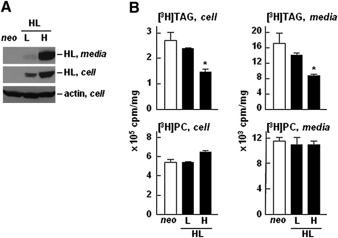
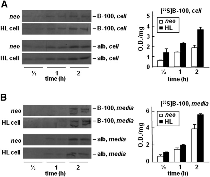
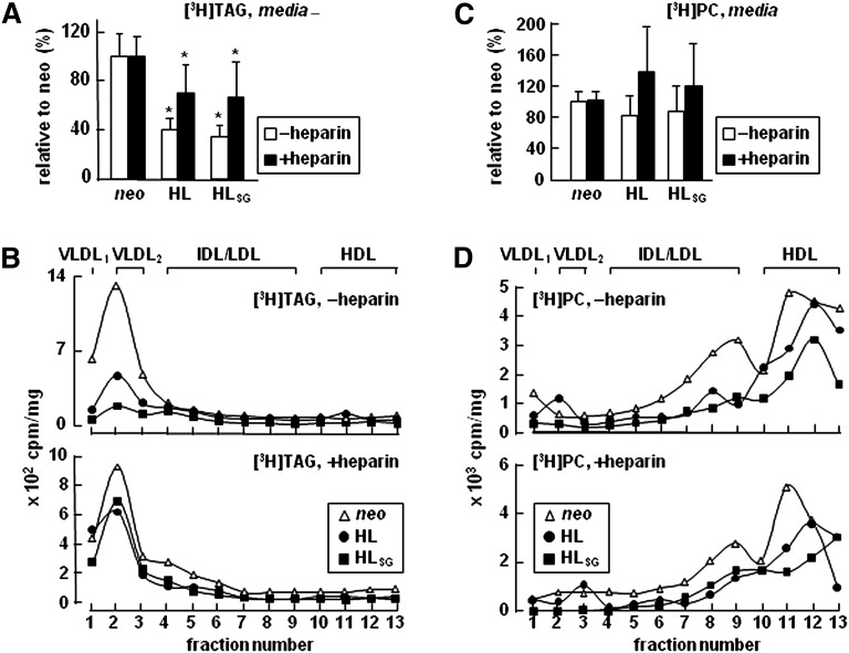
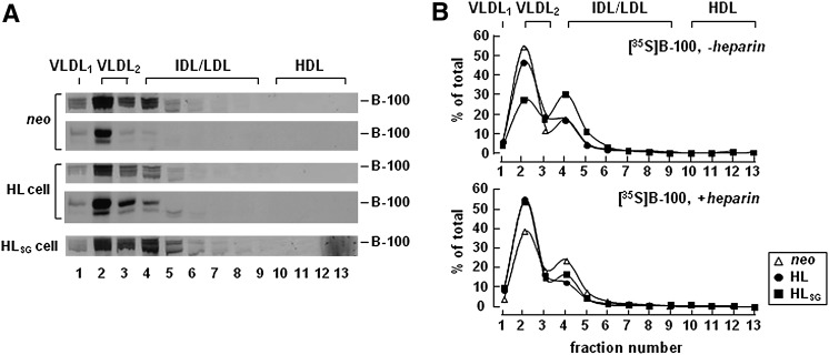
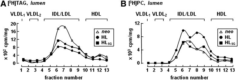
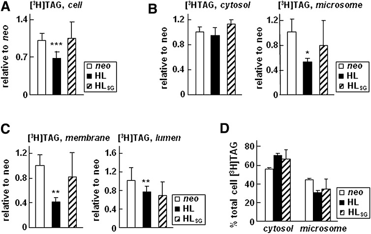
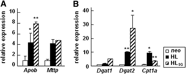
Hepatic lipase (HL) plays a role in the catabolism of apolipoprotein (apo)B-containing lipoproteins through its lipolytic and ligand-binding properties. We describe a potential intracellular role of HL in the assembly and secretion of VLDL. Transient or stable expression of HL in McA-RH7777 cells resulted in decreased (by 40%) incorporation of [(3)H]glycerol into cell-associated and secreted triacylglycerol (TAG) relative to control cells. However, incorporation of [(35)S]methionine/cysteine into cell and medium apoB-100 was not decreased by HL expression. The decreased (3)H-TAG synthesis/secretion in HL expressing cells was not attributable to decreased expression of genes involved in lipogenesis. Fractionation of medium revealed that the decreased [(3)H]TAG from HL expressing cells was mainly attributable to decreased VLDL. Expression of catalytically-inactive HL (HL(SG)) (Ser-145 at the catalytic site was substituted with Gly) in the cells also resulted in decreased secretion of VLDL-[(3)H]TAG. Examination of lumenal contents of microsomes showed a 40% decrease in [(3)H]TAG associated with lumenal lipid droplets in HL or HL(SG) expressing cells as compared with control. The microsomal membrane-associated [(3)H]TAG was decreased by 50% in HL expressing cells but not in HL(SG) expressing cells. Thus, expression of HL, irrespective of its lipolytic function, impairs formation of VLDL precursor [(3)H]TAG in the form of lumenal lipid droplets. These results suggest that HL expression in McA-RH7777 cells result in secretion of [(3)H]TAG-poor VLDL.


Figures

Fig. 1.
Fig. 1.
Expression of HL resulted in decreased synthesis and secretion of [3H]TAG. A: Western blots of recombinant HL expressed in cells and secreted into media (collected from 4 h serum-free DMEM containing 100 U/ml heparin). Actin was used as a loading control. L and H denote low and high levels of HL expressing cells. B: Cells were labeled with [3H]glycerol for 2 h in DMEM containing 20% FBS and 0.4 mM oleate. At the end of the labeling period, lipids were extracted from cells (left panels) and media (right panels), respectively, and separated by TLC. Radioactivity associated with [3H]TAG (top panels) and [3H]PC (bottom panels) was quantified by scintillation counting. The values are expressed as cpm/mg cell protein. * p < 0.05 (Student's t -test of HL vs. neo samples, n = 3).
Fig. 2.
Fig. 2.
Expression of HL does not decrease synthesis or secretion of [35S]apoB-100. Cells were labeled with [35S]methionine/cysteine for 1/2, 1, and 2 h in the presence of 20% FBS and 0.4 mM oleate. At indicated times, apoB-100 and albumin (alb) were recovered from the cells (A) and media (B), respectively, by immunoprecipitation, resolved by SDS-PAGE, and visualized by fluorography (left panels). The intensity of [35S]apoB-100 bands was quantified by scanning densitometry (right panels). Data are presented as the average of two samples (densitometry units/mg cell protein). Repetition of the experiment yielded similar results.
Fig. 3.
Fig. 3.
Decreased secretion of [3H]TAG as VLDL upon HL expression, irrespective of HL catalytic activity. A: Cells were labeled with [3H]glycerol for 2 h in DMEM containing 20% FBS and 0.4 mM oleate ± heparin (100 U/ml). At the end of labeling, lipids were extracted from the media and resolved by TLC. Radioactivity associated with [3H]TAG, quantified by scintillation counting, and was expressed as cpm/mg cell protein. Data presented are converted to [3H]TAG secretion from HL or HLSG cells relative to that from neo control cells (set to 100%). * p < 0.05 (Student's t-test of HL or HLSG vs. neo samples; n = 3). B: The 2 h conditioned labeling media were fractionated by cumulative rate flotation into VLDL1, VLDL2, and other lipoproteins. Lipids were extracted from each fraction, resolved by TLC, and quantified for [3H]TAG. Results are presented as cpm per fraction (normalized with cell proteins). C, D: The experiment was performed the same as in A and B, except that [3H]PC associated with cells (C) and fractionated lipoproteins (D) was quantified.
Fig. 4.
Fig. 4.
Expression of HL did not impair [35S]apoB-100 secretion. A: Cells were labeled with [35S]methionine/cysteine for 2 h in the presence of 20% FBS, 0.4 mM oleate, and ± heparin (100 U/ml). The 2 h conditioned labeling media were fractionated by cumulative rate flotation to separate VLDL1, VLDL2, and other lipoproteins. [35S]apoB-100 in each fraction was recovered by immunoprecipitation, resolved by SDS-PAGE, and subjected to fluorography (Fluorograms for fractional [35S]apoB-100 under plus heparin conditions are not shown). Two independent experiments were performed with neo and HL expressing cells. Experiment with HLSG cells was performed once. B: Radioactivity associated with [35S]apoB-100 in each fraction was quantified by scintillation counting. Data are presented as fractional values as percent of total lumenal [35S]apoB-100 under plus and minus heparin conditions.
Fig. 5.
Fig. 5.
Decreased formation of [3H]TAG-rich lumenal lipid droplets upon HL expression, irrespective of HL catalytic activity. Cells were labeled with [3H]glycerol for 1 h in the presence of 20% FBS and 0.4 mM oleate. Cells were homogenized and microsomal lumenal contents were obtained as described under Materials and Methods. The lumenal contents were fractionated by cumulative rate flotation to separate VLDL1, VLDL2, and other lipoproteins, and the lipids were extracted from each fraction and separated by TLC. The radioactivity associated with [3H]TAG (A) and [3H]PC (B) in each fraction was quantified by scintillation counting. Results are presented as cpm per fraction (normalized with cell proteins).
Fig. 6.
Fig. 6.
Catalytic function of HL is required to decrease the microsomal membrane-associated [3H]TAG. Cell expressing HL, HLSG, or neo control cells were metabolically labeled for 1 or 2 h in DMEM containing 20% FBS and 0.4 mM oleate. A: Total cell-associated [3H]TAG at the end of 2 h labeling. B: [3H]TAG presented in cytosol and microsomes at the end of 1 h labeling. C: [3H]TAG presented between microsomal membrane and microsomal lumen at the end of 1 h labeling. The radioactivity associated with [3H]TAG in A–C were converted to cpm/mg of cell protein, and data are presented as relative values with respect to that of neo control (set to 1). D: Distribution of [3H]TAG (at the end of 1 h labeling) between cytosol and total microsomes. Data are presented as percent of total [3H]TAG. *** p < 0.001; ** p < 0.01; * p < 0.05 (Student's t-test of HL or HLSG vs. neo control samples; n = 4 for neo and HL, and n = 2 for HLSG).
Fig. 7.
Fig. 7.
Relative abundance of mRNA of genes involved in VLDL synthesis or β-oxidation. Total RNAs were extracted from indicated cells and real-time RT PCR analysis was performed on genes encoding apoB-100 (Apob) and MTP (Mttp) (A), DGAT1 (Dgat1), DGAT2 (Dgat2), and carnitine palmitoyl transferase (Cpt1a) (B). Each value is normalized to cyclophilin A (Ppia) and expressed as relative to the level in neo cells. ** p < 0.01; * p < 0.05 (Student's t -test of HL or HLSG vs. neo samples; n = 3).

Similar articles

Cited by

References

    1. Martin G. A., Busch S. J., Meredith G. D., Cardin A. D., Blankenship D. T., Mao S. J., Rechtin A. E., Woods C. W., Racke M. M., Schafer M. P., et al. 1988. Isolation and cDNA sequence of human postheparin plasma hepatic triglyceride lipase. J. Biol. Chem. 263: 10907–10914. - PubMed
    1. Sanan D. A., Fan J., Bensadoun A., Taylor J. M. 1997. Hepatic lipase is abundant on both hepatocyte and endothelial cell surfaces in the liver. J. Lipid Res. 38: 1002–1013. - PubMed
    1. Yu W., Hill J. S. 2006. Mapping the heparin-binding domain of human hepatic lipase. Biochem. Biophys. Res. Commun. 343: 659–665. - PubMed
    1. Stahnke G., Sprengel R., Augustin J., Will H. 1987. Human hepatic triglyceride lipase: cDNA cloning, amino acid sequence and expression in a cultured cell line. Differentiation. 35: 45–52. - PubMed
    1. McCoy M. G., Sun G. S., Marchadier D., Maugeais C., Glick J. M., Rader D. J. 2002. Characterization of the lipolytic activity of endothelial lipase. J. Lipid Res. 43: 921–929. - PubMed

Publication types

LinkOut - more resources