. 2011 Jan 18;108(3):1116-21.
doi: 10.1073/pnas.1018224108. Epub 2010 Dec 28.

Immature T-cell clustering and efficient differentiation require the polarity protein Scribble

Affiliations

Immature T-cell clustering and efficient differentiation require the polarity protein Scribble

Kelly A Pike et al. Proc Natl Acad Sci U S A. .

Abstract

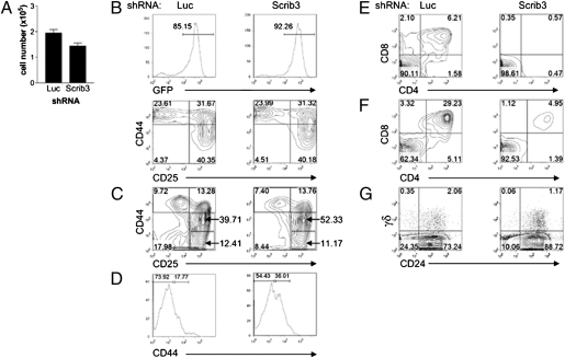
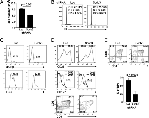
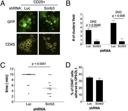
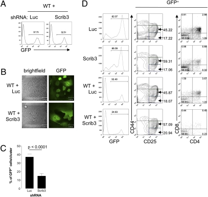
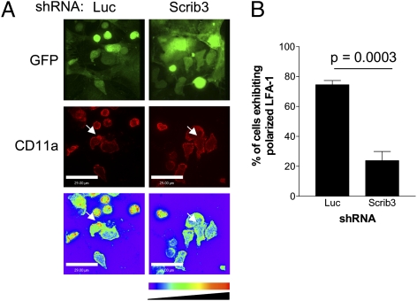
T-cell polarization is required for cell migration and cell-cell interactions, cellular behaviors crucial for lymphocyte differentiation. Despite expression of the epithelial polarity network in T cells, neither its contribution to thymocyte polarity nor its requirement during development is known. We report here that depletion of the polarity protein Scribble in hematopoietic progenitor cells results in inefficient T-cell development characterized by a partial developmental block during the early double-negative (DN) stage of differentiation. Scribble-depleted hematopoietic progenitor cells exhibit a delayed transition into late CD44(lo/-)CD25(+) DN3 cells, evidenced by the accumulation of early CD44(int)CD25(+) DN3 cells. As a consequence, a limited cellular expansion and a reduced frequency of intracellular T-cell receptor β-positive DN3 cells are observed among Scribble-deficient differentiating T cells. Moreover, whereas purified Scribble-depleted DN2 and DN3 cells do not exhibit compromised spontaneous motility, T-cell clustering and prolonged homotypic interactions among such cells are reduced. This deficiency correlates with a lack of polarization of the integrin LFA-1 during T-cell migration or on the initiation of T-cell-T-cell interactions. Scribble is therefore a critical contributor to the clustering of immature T cells, an event shown here to be necessary for efficient developmental progression.


Conflict of interest statement

The authors declare no conflict of interest.

Figures

Fig. 1.
Fig. 1.
Depletion of Scribble gene expression delays T-cell development. (A) Histogram of coculture cellularity on day 5 of culture. The mean ± SEM for culture conditions grown in triplicate are representative of at least three independent experiments. (B and C) The developmental progression of T cells was assessed on day 5 (B) and day 13 (C) of coculture. Representative contour plots are gated on GFP+CD45+ cells and are representative of five independent experiments. (D) After 5–7 d of OP9-DL1 coculture, DN2 (CD45+CD44highCD25+) cells were isolated by cell sorting and plated on fresh OP9-DL1 cells for 5 d. Representative histograms show surface expression levels of CD44 on GFP+CD45+CD25+ cells. Data are representative of four independent experiments. (E) On day 13 of coculture, harvested cells were analyzed for surface expression of CD4 and CD8. Representative contour plots of five independent experiments are shown. (F) After 6 d of culture, cells were harvested and reseeded in the presence of reduced IL-7 concentrations. The contour plots displaying CD4 and CD8 expression are representative of three independent experiments conducted on day 12. (G) Differentiation of γδ T cells was examined on day 11 of coculture. Representative contour plots are gated on GFP+ cells. Numbers in quadrants indicate the percentage of cells within each quadrant.
Fig. 2.
Fig. 2.
Depletion of Scribble gene expression delays the DN3–DP transition. (A) FACS-sorted late DN3 (CD44lo/−CD25+) cells were cultured for 6 d. A representative histogram of culture cellularity is shown. Data represent the mean ± SEM of cultures grown in triplicate, repeated in at least three independent experiments. (B) FACS-sorted late DN3 cells cultured for an additional 3 d were analyzed for cellular DNA content by propidium iodide staining. (C) Histograms of iTCRβ in late DN3 cells, along with an analysis of forward scatter of the corresponding iTCRβ+ late DN3 cells. The histograms are gated on GFP+CD44lo/−CD25+ cells and are representative of at least five independent experiments. (D) Phenotyping of FACS-sorted late DN3 cells after 6 d of coculture. Expression of CD25 was examined on GFP+CD44lo/− cells, CD127 expression was examined on both DN3 and DN4 cells, and finally CD4+CD8+ coexpression among GFP+CD45+ cells was determined. Contour plots and histograms are representative of eight independent experiments. (E) Representative CD4/8 FACS profiles, gated on GFP+ cells, of individual 2-deoxyguanosine–treated thymic lobes reconstituted with Luc KD or Scrib3 KD FTs. A graphic representation of the percentage of DP cells among GFP+ cells in four or five individual thymic lobes within one experiment is shown. Data are representative of three independent experiments. All numbers within FACS profiles correspond to the percentage of cells within each gate, quadrant, or region.
Fig. 3.
Fig. 3.
T-cell–T-cell clustering is limited in the absence of Scribble. (A) Cluster formation among FACS-purified GFP+CD25+ cells adhered to OP9-DL1 cells. (B) Quantification of clusters formed by FACS-sorted DN2 and DN3 cells. The number of clusters within the center field of view is presented. Data represent the mean ± SEM of cultures grown in triplicate, repeated in three independent experiments. (C) Motility of Luc KD and Scrib3 KD immature T cells. Data show the time period during which a cluster remained intact, with each solid circle (Luc KD) or triangle (Scrib3 KD) representing an imaged cluster. The solid bar represents the mean time of cluster maintenance. (D) Scrib3 and Luc KD cells, previously sorted as CD44highCD25+, were plated on OP9-DL1 cells and allowed to adhere for 16 h. Suspension and adherent cells were counted by flow cytometry after staining for CD45. The average percentage of adherent CD45+ cells from all experiments is shown.
Fig. 4.
Fig. 4.
The presence of WT T cells does not rescue clustering or efficient T-cell development. (A) FACS-sorted Scrib3 KD and Luc KD GFP+c-Kit+Sca-1+ HPCs were cultured in the presence of threefold excess of uninfected FACS-sorted GFPc-Kit+Sca-1+ HPCs. Representative histograms of 10,000 events are shown. (B) Bright-field images of mixed cocultures compared with images of GFP fluorescence. (C) The proportion of GFP+ cells within 40 analyzed clusters in Scrib3 KD + WT or Luc KD + WT cocultures between day 8 and day 10 of culture. (D) Cultures were harvested on day 12 of coculture and phenotyped for developmental progression. The contour plots of CD44 and CD25 expression and dot plots of CD4 and CD8 expression shown are representative of three independent experiments. All plots are gated on GFP+ cells, and numbers indicate the percentage of cells within indicated gates or quadrants.
Fig. 5.
Fig. 5.
Scribble is required for LFA-1 polarization on immature T cells. (A) Localization of LFA-1 on immature T cells was visualized by fixation and surface staining of cocultures by staining for CD11a. Representative extended-focus images of polarized CD25+ immature T cells of either GFP or LFA-1 (Cy-3) are shown. A color look-up table was applied to all images of LFA-1 staining. White arrows identify sites of accumulated LFA-1. (B) The incidence of polarized LFA-1 among >100 cells from three independent experiments.

References

    1. Dow LE, Humbert PO. Polarity regulators and the control of epithelial architecture, cell migration, and tumorigenesis. Int Rev Cytol. 2007;262:253–302. - PubMed
    1. Ladi E, Yin X, Chtanova T, Robey EA. Thymic microenvironments for T cell differentiation and selection. Nat Immunol. 2006;7:338–343. - PubMed
    1. Takahama Y. Journey through the thymus: Stromal guides for T-cell development and selection. Nat Rev Immunol. 2006;6:127–135. - PubMed
    1. Lind EF, Prockop SE, Porritt HE, Petrie HT. Mapping precursor movement through the postnatal thymus reveals specific microenvironments supporting defined stages of early lymphoid development. J Exp Med. 2001;194:127–134. - PMC - PubMed
    1. Porritt HE, Gordon K, Petrie HT. Kinetics of steady-state differentiation and mapping of intrathymic-signaling environments by stem cell transplantation in nonirradiated mice. J Exp Med. 2003;198:957–962. - PMC - PubMed

Publication types

MeSH terms

Substances