. 2011 Mar;79(3):1124-33.
doi: 10.1128/IAI.00658-10. Epub 2010 Dec 28.

Differential microbicidal effects of human histone proteins H2A and H2B on Leishmania promastigotes and amastigotes

Affiliations

Differential microbicidal effects of human histone proteins H2A and H2B on Leishmania promastigotes and amastigotes

Yingwei Wang et al. Infect Immun. 2011 Mar.

Abstract

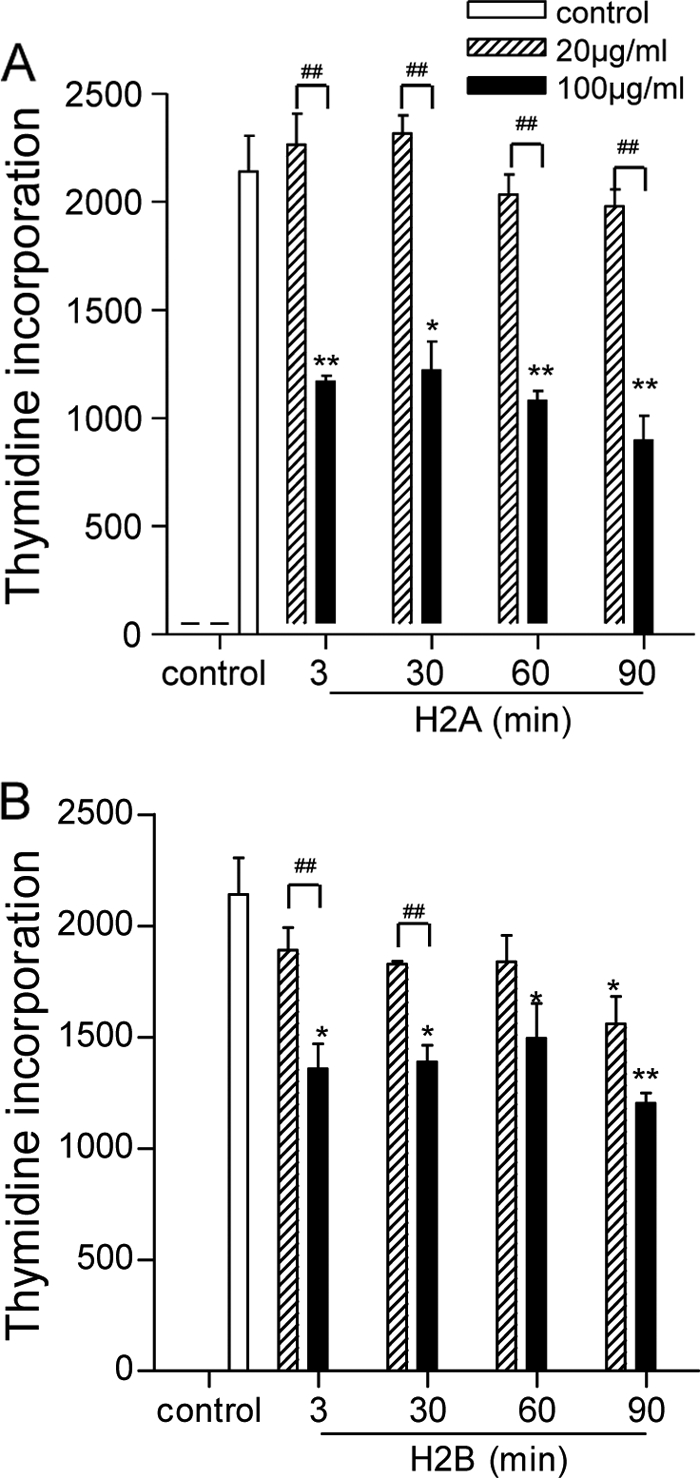
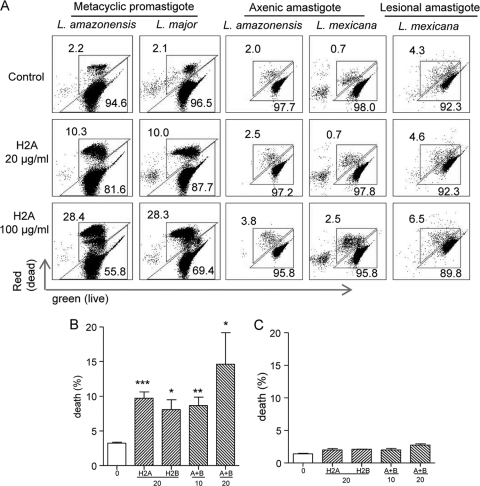
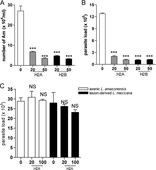
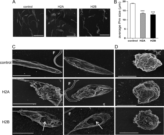
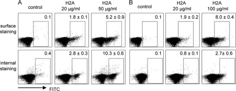
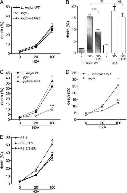
Recent studies have shown that histone proteins can act as antimicrobial peptides in host defense against extracellular bacteria, fungi, and Leishmania promastigotes. In this study, we used human recombinant histone proteins to further study their leishmaniacidal effects and the underlying mechanisms. We found that the histones H2A and H2B (but not H1(0)) could directly and efficiently kill promastigotes of Leishmania amazonensis, L. major, L. braziliensis, and L. mexicana in a treatment dose-dependent manner. Scanning electron microscopy revealed surface disruption of histone-treated promastigotes. More importantly, the preexposure of promastigotes to histone proteins markedly decreased the infectivity of promastigotes to murine macrophages (Mφs) in vitro. However, axenic and lesion-derived amastigotes of L. amazonensis and L. mexicana were relatively resistant to histone treatment, which correlated with the low levels of intracellular H2A in treated amastigotes. To understand the mechanisms underlying these differential responses, we investigated the role of promastigote surface molecules in histone-mediated killing. Compared with the corresponding controls, transgenic L. amazonensis promastigotes expressing lower levels of surface gp63 proteins were more susceptible to histone H2A, while L. major and L. mexicana promastigotes with targeted deletion of the lipophosphoglycan 2 (lpg2) gene (but not the lpg1 gene) were more resistant to histone H2A. We discuss the influence of promastigote major surface molecules in the leishmaniacidal effect of histone proteins. This study provides new information on host innate immunity to different developmental stages of Leishmania parasites.


Figures

FIG. 1.
FIG. 1.
Suppressive effects of histone H2A and H2B on parasite proliferation. L. amazonensis promastigotes (2.5 × 10⁶ in 50 μl PBS) were treated with 20 or 100 μg/ml of histone H2A (A) or H2B (B) at 23°C for indicated time periods, after which complete Schneider's medium was added. After 24 h of incubation, [3H]thymidine was added for an additional 18 h of incubation. Incorporated [3H]thymidine was measured and presented as counts per minute (cpm). Data are pooled from two independent repeats and shown as the means ± the standard errors. * (P < 0.05) and ** (P < 0.01) indicate statistically significant differences between the control and treated groups. # (P < 0.05) and ## (P < 0.01) indicate the statistical differences between the marked groups.
FIG. 2.
FIG. 2.
Direct killing of Leishmania promastigotes, but not amastigotes, by histone proteins. Promastigotes of L. amazonensis (A and B), L. major (C), and L. braziliensis (D) were treated with 20 to 200 μg/of of histone H2A, H2B, or H10 for 30 min and then stained with calcein AM and ethidium homodimer for evaluating live (green) versus dead (red) parasites, respectively. Shown are representative results from three independent repeats, and data are presented as mean survival rates (%) ± standard errors. ** (P < 0.01) and *** (P < 0.001) indicate statistically significant differences between the control and treated groups. # (P < 0.05), ## (P < 0.01), and ### (P < 0.001) indicate the statistical differences between the marked groups.
FIG. 3.
FIG. 3.
Analysis of the death rates of histone-treated parasites by flow cytometry. (A) Metacyclic promastigotes of L. major and L. amazonensis were purified by using Ficoll gradient centrifugation and treated with an indicated concentration of histone H2A. Axenic amastigotes of L. amazonensis and L. mexicana, as well as lesion-derived amastigotes of L. mexicana, were treated similarly. Parasites were stained with calcein AM and ethidium homodimer and analyzed on a C6 flow cytometer. The numbers represent the percentages of dead (top left numbers) and live (bottom right numbers) parasites. (B and C) Promastigotes (B) and axenic amastigotes (C) of L. amazonensis were treated with histone H2A or H2B alone or in combination at indicated concentrations (μg/ml) and analyzed as described for panel A. Shown are representative results from two independent repeats, and data are presented as mean death rates (%) ± standard errors. * (P < 0.05), ** (P < 0.01), and *** (P < 0.001) indicate statistically significant differences between the control and treated groups.
FIG. 4.
FIG. 4.
Mφ infection with histone-treated L. amazonensis parasites. (A and B) L. amazonensis promastigotes were treated with an indicated concentration of H2A or H2B for 30 min and used to infect Mφs (at a 5:1 parasite-to-cell ratio). The number of intracellular parasites was determined at 72 h by direct counting under a hemocytometer (A) or via real-time PCR analysis (B). (C) Mφs were infected with histone-pretreated axenic amastigotes of L. amazonensis (at a 5:1 parasite-to-cell ratio) or lesion-derived amastigotes of L. mexicana (at a 1:2 parasite-to-cell ratio) for 48 h. Parasite loads were determined by real-time PCR analysis. Shown are means ± standard errors. *** (P < 0.001) indicates statistically significant differences between the control groups and histone-treated groups, and NS means no significance.
FIG. 5.
FIG. 5.
Morphological and ultrastructural changes of histone-treated parasites in scanning electron microscopy (SEM). L. amazonensis promastigotes were treated with histone H2A or H2B (100 μg/ml) for 30 min, and then samples were immediately fixed for SEM analysis. (A) Representative SEM images for control and treated parasites. Bars, 20 μm. (B) Data are shown as the average lengths of promastigotes in the control and histone-treated groups. Shown are means ± standard errors. *** (P < 0.001) indicates statistically significant differences between the control and treated groups. (C and D) L. amazonensis promastigotes (C) and axenic amastigotes (D) were left untreated or treated with histone H2A or H2B (100 μg/ml) for 30 min. Samples were fixed for SEM analysis, and images were taken at ×15,000 to ×20,000 magnification. Bars, 3 μm (C) and 2 μm (D). Arrows points to variable sizes of holes on the surface of promastigotes. F, flagellum.
FIG. 6.
FIG. 6.
Surface and internal binding of histone proteins on Leishmania parasites. L. amazonensis promastigotes (A) or axenic amastigotes (B) were treated with the indicated concentration of histone H2A for 30 min. After washing, surface-bound H2A was stained with a mouse MAb specific to human histone H2A and then with an FITC-conjugated anti-mouse IgG1 antibody. For detecting internalized H2A, parasites were permeabilized prior to and during antibody staining. Parasites were analyzed on a C6 flow cytometer. The numbers in the boxes indicate the percentages of positively stained parasites, and data are presented as means (%) ± standard errors. Shown are representative results from two independent repeats.
FIG. 7.
FIG. 7.
Effects of LPG and gp63 on histone-mediated promastigote killing. (A) Wild-type (WT), lpg1, and lpg1/+LPG1 promastigotes of L. major were treated with the indicated concentrations (μg/ml) of histone H2A or H2B and then stained with calcein AM and ethidium homodimer, respectively. The stained parasites were then analyzed on a C6 flow cytometer. (B) Histone H2A (50 μg/ml) was preincubated with purified L. major LPG (50 μg/ml) for 30 min prior to parasite treatment and death analysis. (C) WT, lpg2, and lpg2/+LPG2 promastigotes of L. major were treated with indicated concentrations (μg/ml) of histone H2A and analyzed for death rates. (D) WT and lpg2 promastigotes of L. mexicana were treated with the indicated concentrations of histone H2A and analyzed for death. (E) WT (P6.5), P6.5/1.9, and P6.5/1.9R promastigotes of L. amazonensis were treated with the indicated concentrations of H2A and analyzed for death. Shown are the means ± the standard errors. * (P < 0.05), ** (P < 0.01), and *** (P < 0.001) indicate statistically significant differences between histone-treated groups. NS means no significance among these groups.

Similar articles

Cited by

References

    1. Appelberg, R. 2007. Neutrophils and intracellular pathogens: beyond phagocytosis and killing. Trends Microbiol. 15:87-92. - PubMed
    1. Bahr, V., et al. 1993. Expression of lipophosphoglycan, high-molecular weight phosphoglycan and glycoprotein 63 in promastigotes and amastigotes of Leishmania mexicana. Mol. Biochem. Parasitol. 58:107-121. - PubMed
    1. Brinkmann, V., et al. 2004. Neutrophil extracellular traps kill bacteria. Science 303:1532-1535. - PubMed
    1. Chan, D. I., E. J. Prenner, and H. J. Vogel. 2006. Tryptophan- and arginine-rich antimicrobial peptides: structures and mechanisms of action. Biochim. Biophys. Acta 1758:1184-1202. - PubMed
    1. Chen, D. Q., et al. 2000. Episomal expression of specific sense and antisense mRNAs in Leishmania amazonensis: modulation of gp63 level in promastigotes and their infection of macrophages in vitro. Infect. Immun. 68:80-86. - PMC - PubMed

Publication types

MeSH terms

LinkOut - more resources