. 2011 Feb 1;71(3):964-75.
doi: 10.1158/0008-5472.CAN-10-3172. Epub 2010 Dec 28.

Disruption of a Sirt1-dependent autophagy checkpoint in the prostate results in prostatic intraepithelial neoplasia lesion formation

Affiliations

Disruption of a Sirt1-dependent autophagy checkpoint in the prostate results in prostatic intraepithelial neoplasia lesion formation

Michael J Powell et al. Cancer Res. .

Abstract

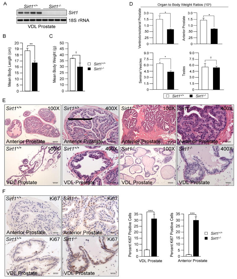
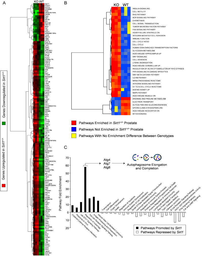
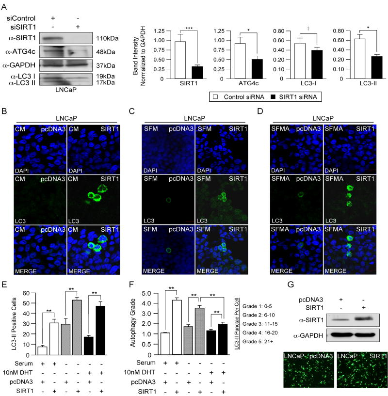
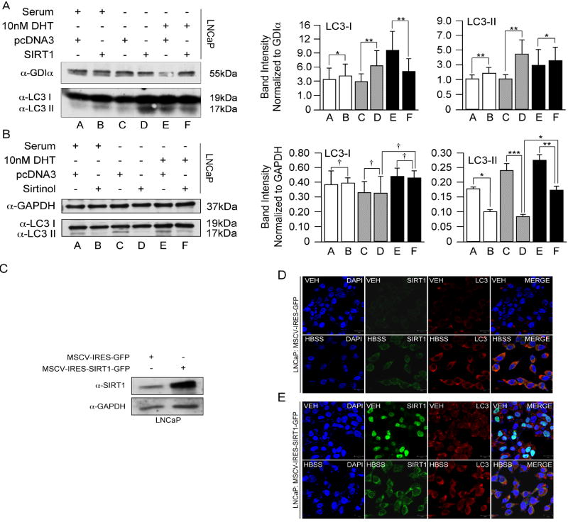
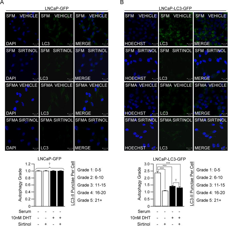
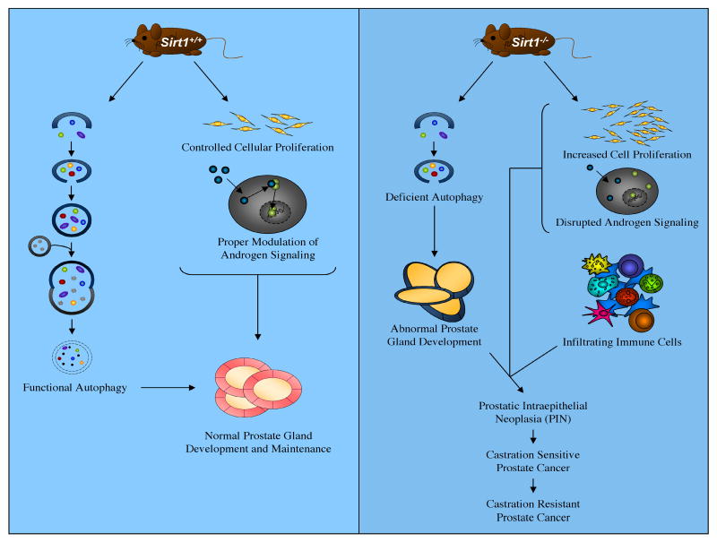
The Sirtuin family of proteins (SIRT) encode a group of evolutionarily conserved, NAD-dependent histone deacetylases, involved in many biological pathways. SIRT1, the human homologue of the yeast Silent Information Regulator 2 (Sir2) gene, deacetylates histones, p300, p53, and the androgen receptor. Autophagy is required for the degradation of damaged organelles and long-lived proteins, as well as for the development of glands such as the breast and prostate. Herein, homozygous deletion of the Sirt1 gene in mice resulted in prostatic intraepithelial neoplasia (PIN) associated with reduced autophagy. Genome-wide gene expression analysis of Sirt1(-/-) prostates demonstrated that endogenous Sirt1 repressed androgen responsive gene expression and induced autophagy in the prostate. Sirt1 induction of autophagy occurred at the level of autophagosome maturation and completion in cultured prostate cancer cells. These studies provide novel evidence for a checkpoint function of Sirt1 in the development of PIN and further highlight a role for SIRT1 as a tumor suppressor in the prostate.


Conflict of interest statement

The authors declare no conflict of interest as they pertain to this manuscript.

Figures

Figure 1
Figure 1. Sirt1 Deletion Alters Androgen-Responsive Tissue Development
(A) RT-PCR using ventrodorsolateral prostate RNA from Sirt1+/+ and Sirt1-/- mice. (B) Mean body length, (C) mass, and (D) select tissue weights normalized to total mass (see also Fig. S1A). (E) Hematoxylin and Eosin (H&E) staining of prostate glands from Sirt1+/+and Sirt1-/- mice (see also Fid. S1B). (F) Ki67 staining of Sirt1+/+ and Sirt1-/- prostates. All data are mean ± standard error of the mean (SEM) and represent n=5/genotype. P values were determined by student's t-test (*P<0.05, **P<0.01, ***P<0.001, † denotes no statistical significance).
Figure 2
Figure 2. Sirt1 Governs Vital Biological Pathways in the Prostate
(A) Treeview of microarray data generated from Sirt1+/+ and Sirt1-/- ventrodorsolateral prostates. (B) Heatmap displaying pathways affected by Sirt1 in the prostate as determined by the Analysis of Sample Set Enrichment Scores (ASSESS) database (see also Fig. S1C, Tables S1-S4). (C) Graphical schematic highlighting Sirt1 affected pathways in the prostate as revealed by the Database for Annotation, Visualization and Integrated Discovery (DAVID). Also illustrated is Sirt1's ability to affect a subset of autophagy related genes (Atg's) involved in autophagosome maturation and completion.
Figure 3
Figure 3. SIRT1 Antagonizes AR-Mediated Inhibition of Autophagy
(A) Western blot for SIRT1, ATG4c, and LC3 following SIRT1 siRNA treatment in LNCaP cells. All data are representative of triplicate experiments. Statistical significance was determined by Student's t-test (*P<0.05, ***P<0.001, † denotes no statistical significance). (B-D) LNCaP cells transfected with pcDNA3 or SIRT1 and cultured in complete serum media (CM), serum free media (SFM), or serum free media supplemented with 10nM DHT (SFMA) were subjected to LC3-II immunofluorescence (see also Figs. S2A-S2C). (E,F) Quantitation of autophagic cells based on LC3-II positivity (ANOVA, **P<0.01) and autophagic grade (Mann Whitney U, **P<0.01). All data are representative of triplicate experiments. Quantitation represents at least 100 cells counted and scored per treatment. (G) SIRT1 Western blot following co-transfection with pcDNA3 vector control or SIRT1 and GFP (micrographs confirm GFP-positive cells post-transfection).
Figure 4
Figure 4. SIRT1 Promotes Autophagy in Prostate Cancer Cells
(A,B) LC3 Western blots in LNCaP cells following SIRT1 over-expression or inhibition via Sirtinol (30μM) in various media conditions. (C) Western blot verifying SIRT1 abundance in LNCaP cells transduced with MSCV-IRES-GFP or MSCV-IRES-SIRT1-GFP retroviruses. (D,E) SIRT1 and LC3 dual-immunofluorescence of transduced LNCaPs in the presence or absence of autophagic stimuli (HBSS) (see also Fig. S7). All data are representative of triplicate experiments. Statistical significance was measured by ANOVA (*P<0.05, **P<0.01, ***P<0.0001, † denotes no statistical significance).
Figure 5
Figure 5. SIRT1 Enhances LC3 Cleavage and Autophagy Regardless of Media Conditions
(A,B) LNCaP cells stably expressing GFP (LNCaP-GFP) or a GFP-tagged, pre-cleaved version of LC3 (LNCaP-LC3-GFP) transfected with SIRT1 or pcDNA3 vector control. (C,D) LNCaP-GFP and LNCaP-LC3-GFP cells transfected with pcDNA3 or SIRT1 in the presence and absence of DHT. Quantitation of triplicate experiments was performed based on autophagic grade and graphed adjacent to micrographs (Mann Whitney U, **P<0.01, ***P<0.001, † denotes no statistical significance).
Figure 6
Figure 6. Chemical Inhibition of SIRT1 Abolishes Autophagy in Prostate Cancer Cells
(A,B) LNCaP-GFP and LNCaP-LC3-GFP cells cultured in various media conditions in the presence and absence of Sirtinol (30μM) and analyzed by confocal microscopy for LC3 punctae. Statistical significance of triplicate experiments was measured by Mann Whitney U test (***P<0.001, † denotes no statistical significance).
Figure 7
Figure 7. SIRT1 Promotes Autophagy-Mediated Prostate Development and Health
(A) Proposed model of SIRT1 function in the prostate as it relates to autophagy and prostate homeostasis.

References

    1. Yang T, Fu M, Pestell R, Sauve AA. SIRT1 and endocrine signaling. Trends Endocrinol Metab. 2006;17:186–91. - PubMed
    1. Whittle JR, Powell MJ, Popov VM, Shirley LA, Wang C, Pestell RG. Sirtuins, nuclear hormone receptor acetylation and transcriptional regulation. Trends Endocrinol Metab. 2007;18:356–64. - PubMed
    1. Longo VD, Kennedy BK. Sirtuins in aging and age-related disease. Cell. 2006;126:257–68. - PubMed
    1. Lim CS. SIRT1: tumor promoter or tumor suppressor? Med Hypotheses. 2006;67:341–4. - PubMed
    1. Ota H, Tokunaga E, Chang K, Hikasa M, Iijima K, Eto M, et al. Sirt1 inhibitor, Sirtinol, induces senescence-like growth arrest with attenuated Ras-MAPK signaling in human cancer cells. Oncogene. 2006;25:176–85. - PubMed

Publication types