

Novel bacterial NAD+-dependent DNA ligase inhibitors with broad-spectrum activity and antibacterial efficacy in vivo
- PMID: 21189350
- PMCID: PMC3067096
- DOI: 10.1128/AAC.01181-10
Novel bacterial NAD+-dependent DNA ligase inhibitors with broad-spectrum activity and antibacterial efficacy in vivo
Abstract
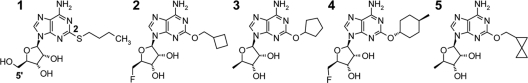
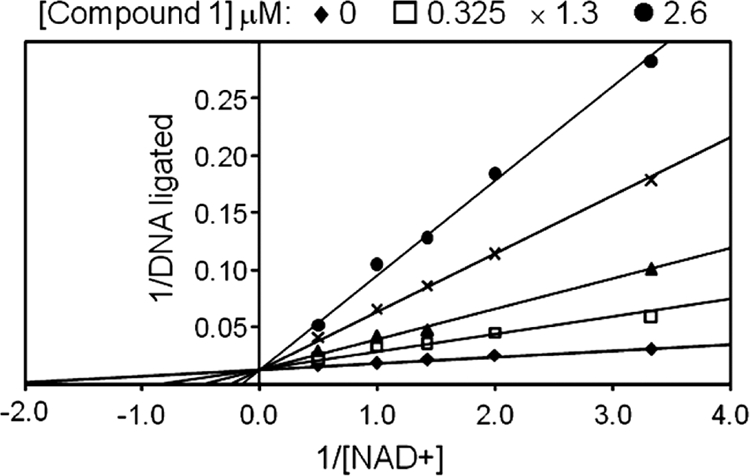
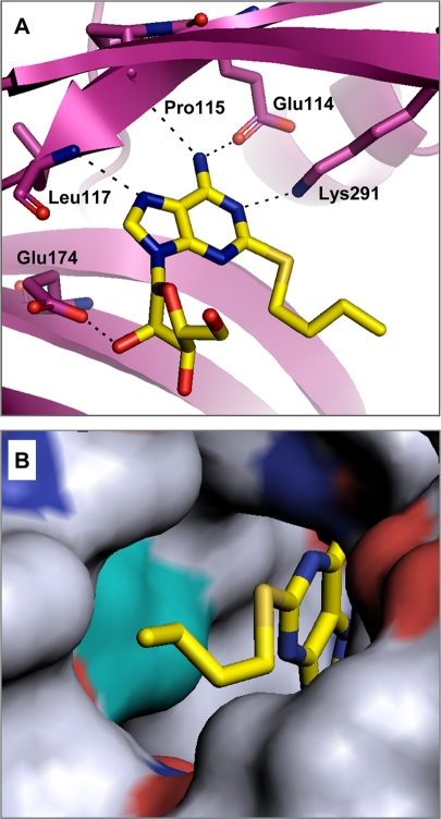
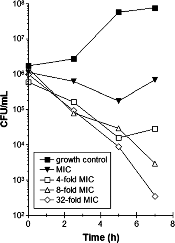
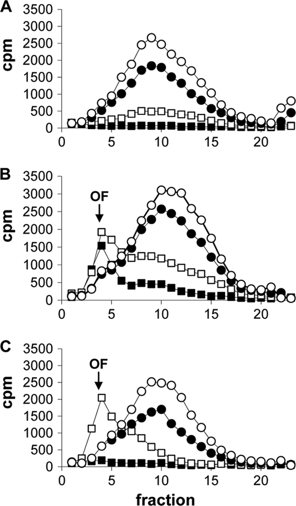
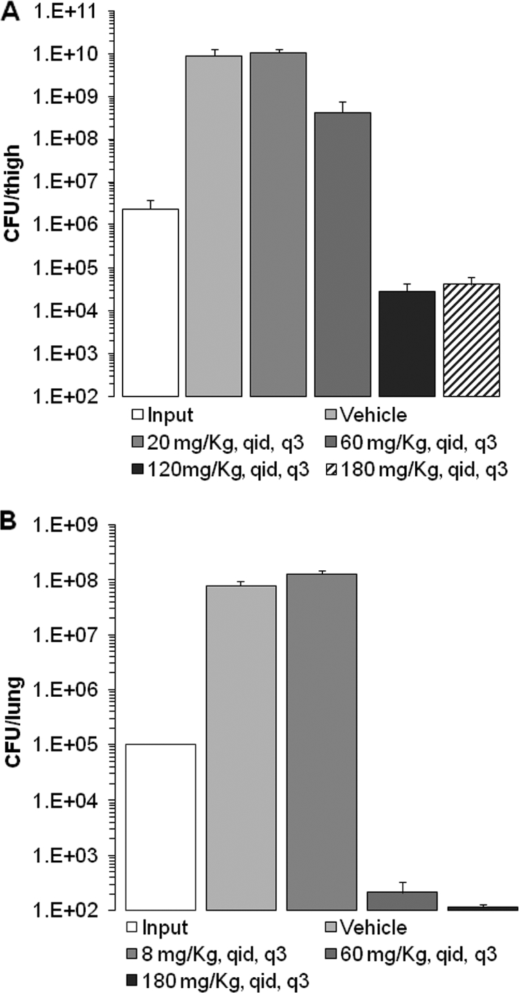
DNA ligases are indispensable enzymes playing a critical role in DNA replication, recombination, and repair in all living organisms. Bacterial NAD+-dependent DNA ligase (LigA) was evaluated for its potential as a broad-spectrum antibacterial target. A novel class of substituted adenosine analogs was discovered by target-based high-throughput screening (HTS), and these compounds were optimized to render them more effective and selective inhibitors of LigA. The adenosine analogs inhibited the LigA activities of Escherichia coli, Haemophilus influenzae, Mycoplasma pneumoniae, Streptococcus pneumoniae, and Staphylococcus aureus, with inhibitory activities in the nanomolar range. They were selective for bacterial NAD+-dependent DNA ligases, showing no inhibitory activity against ATP-dependent human DNA ligase 1 or bacteriophage T4 ligase. Enzyme kinetic measurements demonstrated that the compounds bind competitively with NAD+. X-ray crystallography demonstrated that the adenosine analogs bind in the AMP-binding pocket of the LigA adenylation domain. Antibacterial activity was observed against pathogenic Gram-positive and atypical bacteria, such as S. aureus, S. pneumoniae, Streptococcus pyogenes, and M. pneumoniae, as well as against Gram-negative pathogens, such as H. influenzae and Moraxella catarrhalis. The mode of action was verified using recombinant strains with altered LigA expression, an Okazaki fragment accumulation assay, and the isolation of resistant strains with ligA mutations. In vivo efficacy was demonstrated in a murine S. aureus thigh infection model and a murine S. pneumoniae lung infection model. Treatment with the adenosine analogs reduced the bacterial burden (expressed in CFU) in the corresponding infected organ tissue as much as 1,000-fold, thus validating LigA as a target for antibacterial therapy.
Figures
References
-
- Azoulay-Dupuis, E., et al. 1991. Antipneumococcal activity of ciprofloxacin, ofloxacin and temafloxacin in an experimental mouse pneumonia model at various stages of the disease. J. Infect. Dis. 163:319-324. - PubMed
-
- Balani, S. K., et al. 2004. Effective dosing regimen of 1-aminobenzotriazole for inhibition of antipyrine clearance in guinea pigs and mice using serial sampling. Drug Metab. Dispos. 32:1092-1095. - PubMed
-
- Benson, E. L., et al. 2004. A high-throughput resonance energy transfer assay for Staphylococcus aureus DNA ligase. Anal. Biochem. 324:298-300. - PubMed
MeSH terms
Substances
LinkOut - more resources
Full Text Sources
Other Literature Sources
Medical
Molecular Biology Databases
Research Materials
Miscellaneous
