. 2011 Jan;52(1):154-61.
doi: 10.2967/jnumed.110.079343.

MRI-assisted PET motion correction for neurologic studies in an integrated MR-PET scanner

Affiliations

MRI-assisted PET motion correction for neurologic studies in an integrated MR-PET scanner

Ciprian Catana et al. J Nucl Med. 2011 Jan.

Abstract

Head motion is difficult to avoid in long PET studies, degrading the image quality and offsetting the benefit of using a high-resolution scanner. As a potential solution in an integrated MR-PET scanner, the simultaneously acquired MRI data can be used for motion tracking. In this work, a novel algorithm for data processing and rigid-body motion correction (MC) for the MRI-compatible BrainPET prototype scanner is described, and proof-of-principle phantom and human studies are presented.

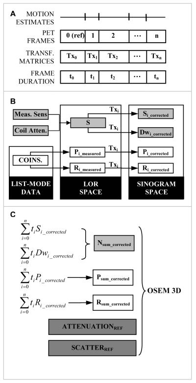
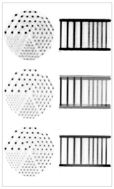
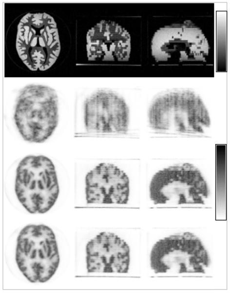
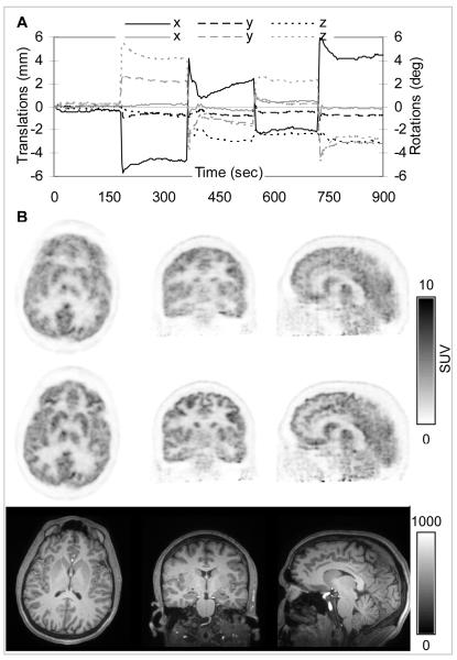
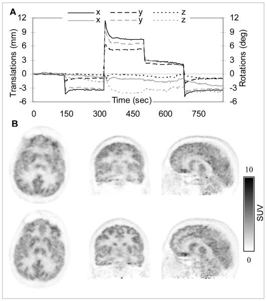
Methods: To account for motion, the PET prompt and random coincidences and sensitivity data for postnormalization were processed in the line-of-response (LOR) space according to the MRI-derived motion estimates. The processing time on the standard BrainPET workstation is approximately 16 s for each motion estimate. After rebinning in the sinogram space, the motion corrected data were summed, and the PET volume was reconstructed using the attenuation and scatter sinograms in the reference position. The accuracy of the MC algorithm was first tested using a Hoffman phantom. Next, human volunteer studies were performed, and motion estimates were obtained using 2 high-temporal-resolution MRI-based motion-tracking techniques.

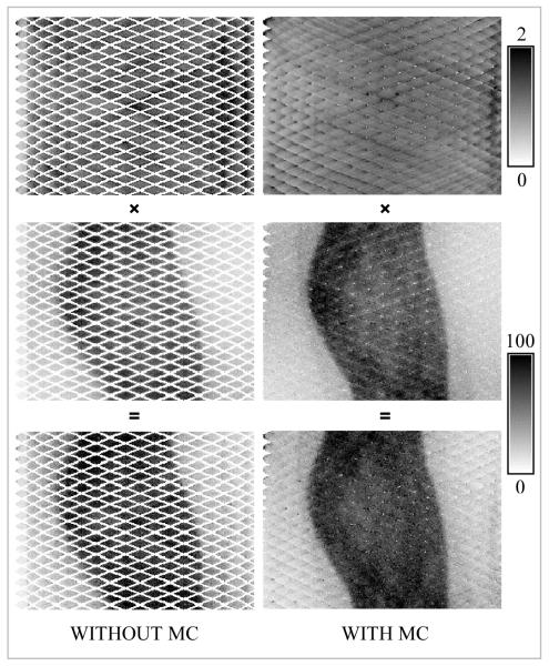
Results: After accounting for the misalignment between the 2 scanners, perfectly coregistered MRI and PET volumes were reproducibly obtained. The MRI output gates inserted into the PET list-mode allow the temporal correlation of the 2 datasets within 0.2 ms. The Hoffman phantom volume reconstructed by processing the PET data in the LOR space was similar to the one obtained by processing the data using the standard methods and applying the MC in the image space, demonstrating the quantitative accuracy of the procedure. In human volunteer studies, motion estimates were obtained from echo planar imaging and cloverleaf navigator sequences every 3 s and 20 ms, respectively. Motion-deblurred PET images, with excellent delineation of specific brain structures, were obtained using these 2 MRI-based estimates.

Conclusion: An MRI-based MC algorithm was implemented for an integrated MR-PET scanner. High-temporal-resolution MRI-derived motion estimates (obtained while simultaneously acquiring anatomic or functional MRI data) can be used for PET MC. An MRI-based MC method has the potential to improve PET image quality, increasing its reliability, reproducibility, and quantitative accuracy, and to benefit many neurologic applications.


Figures

FIGURE 1
FIGURE 1
BrainPET scanner – 3D rendering showing the placement of the detector blocks inside the gantry (left). Transaxial (top right) and axial (bottom right) sections illustrating the gaps between the LSO arrays: 32 in the transaxial plane and 5 in the axial direction.
FIGURE 2
FIGURE 2
Motion compensation algorithm for the BrainPET data workflow: (A) PET data framing based on the motion estimates (derived from MR); (B) LOR-space processing of each individual frame based on the transformation matrices (Tx) and rebinning into the motion corrected sinograms: prompts (P) and smoothed delays (R) coincidences, dwell (Dw) and sensitivity (S); (C) Generation of the motion corrected data by summing the time weighted sinograms and image reconstruction using the standard OP-OSEM 3D algorithm.
FIGURE 3
FIGURE 3
Simultaneously acquired MR-PET data using a Derenzo phantom: representative PET images (top), fused MR-PET before (middle) and after (bottom) accounting for the spatial mismatch between the two scanners. Images in the transaxial and coronal orientations are shown in each case. Note the perfect co-registration between the two volumes after performing the motion correction.
FIGURE 4
FIGURE 4
Representative sinograms for the BrainPET scanner before and after applying spatial transformations. The normalization and the corresponding prompt sinograms from the data acquired with the Hoffman phantom and their product are shown in the top, middle and bottom rows, respectively. Note the empty bins in the data uncorrected for motion. After applying the transformation most of these bins were filled with data in both the prompts as well as the normalization sinograms.
FIGURE 5
FIGURE 5
MR-based MC in a Hoffman phantom using MPRAGE-derived motion estimates: MR images in the reference position (top row), uncorrected PET images (second row), data corrected in the LOR space before image reconstruction (third row) and in the image space after reconstructing each individual frame (forth row). Note the substantial improvement in image quality after MC. Images in the transaxial, coronal and sagittal orientations are shown in each case.
FIGURE 6
FIGURE 6
MR-based MC in a healthy volunteer using fMRI-derived motion estimates: (A) Plot of the motion estimates in the PET reference frame: translations along (black) and rotations about (gray) the three orthogonal axes are shown. (B) PET data reconstructed before (first row) and after motion correction (second row). Note the substantial improvement in the PET image quality after MC. The corresponding MR images are provided as a reference (third row).
FIGURE 7
FIGURE 7
MR-based MC in a healthy volunteer using CLN-derived motion estimates: (A) Plot of the motion estimates in the PET reference frame: translations along (black) and rotations about (gray) the three orthogonal axes are shown. (B) PET data reconstructed before (first row) and after MC (second row). Note the substantial improvement in the PET image quality after MC.

References

    1. Catana C, Procissi D, Wu Y, et al. Simultaneous in vivo positron emission tomography and magnetic resonance imaging. Proc Natl Acad Sci USA. 2008 Mar 11;105(10):3705–3710. - PMC - PubMed
    1. Judenhofer MS, Wehrl HF, Newport DF, et al. Simultaneous PET-MRI: a new approach for functional and morphological imaging. Nat Med. 2008 Apr;14(4):459–465. - PubMed
    1. Lucas AJ, Hawkes RC, Ansorge RE, et al. Development of a combined microPET((R))-MR system. Technol Cancer Res Treat. 2006 Aug;5(4):337–341. - PubMed
    1. Raylman RR, Majewski S, Velan SS, et al. Simultaneous acquisition of magnetic resonance spectroscopy (MRS) data and positron emission tomography (PET) images with a prototype MR-compatible, small animal PET imager. J Magn Reson. 2007 Jun;186(2):305–310. - PMC - PubMed
    1. Schlemmer H-PW, Pichler BJ, Schmand M, et al. Simultaneous MR/PET Imaging of the Human Brain: Feasibility Study. Radiology. 2008 Sep 1;248(3):1028–1035. - PubMed

Publication types