. 2011 Mar 11;286(10):8597-8608.
doi: 10.1074/jbc.M110.186742. Epub 2010 Dec 28.

Molecular basis of Wnt activation via the DIX domain protein Ccd1

Affiliations

Molecular basis of Wnt activation via the DIX domain protein Ccd1

Yi-Tong Liu et al. J Biol Chem. .

Abstract

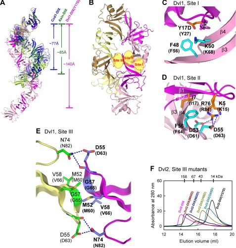
The Wnt signaling plays pivotal roles in embryogenesis and cancer, and the three DIX domain-containing proteins, Dvl, Axin, and Ccd1, play distinct roles in the initiation and regulation of canonical Wnt signaling. Overexpressed Dvl has a tendency to form large polymers in a cytoplasmic punctate pattern, whereas the biologically active Dvl in fact forms low molecular weight oligomers. The molecular basis for how the polymeric sizes of Dvl proteins are controlled upon Wnt signaling remains unclear. Here we show that Ccd1 up-regulates canonical Wnt signaling via acting synergistically with Dvl. We determined the crystal structures of wild type Ccd1-DIX and mutant Dvl1-DIX(Y17D), which pack into "head-to-tail" helical filaments. Structural analyses reveal two sites crucial for intra-filament homo- and hetero-interaction and a third site for inter-filament homo-assembly. Systematic mutagenesis studies identified critical residues from all three sites required for Dvl homo-oligomerization, puncta formation, and stimulation of Wnt signaling. Remarkably, Ccd1 forms a hetero-complex with Dvl through the "head" of Dvl-DIX and the "tail" of Ccd1-DIX, depolymerizes Dvl homo-assembly, and thereby controls the size of Dvl polymer. These data together suggest a molecular mechanism for Ccd1-mediated Wnt activation in that Ccd1 converts latent polymeric Dvl to a biologically active oligomer(s).


Figures

FIGURE 1.
FIGURE 1.
Ccd1 activates canonical Wnt pathway through Dvl. A, knockdown of endogenous Ccd1 attenuates Wnt-3a mediated T-cell factor transcriptional activity. HEK293T cells were transfected with Wnt-3a (0 or 2 ng) and pSUPER-Ccd1 (0 or 30 ng) as indicated. Some 36 h later the activation of Wnt signaling was quantified by luciferase activity. Asterisks denote p < 0.001 when compared with the corresponding samples only transfected with Wnt-3a. B, knockdown of endogenous Dvl suppresses Ccd1 function. HEK293T cells were cotransfected with 100 nm Dvl2/3 siRNAs together with Wnt-3a (2 ng) or Ccd1 (10 ng). *, p < 0.05; **, p < 0.005 compared with the corresponding samples transfected with control siRNAs. Ctr, control. C, synergistic effects of Dvl and Ccd1 on Wnt activation. Some 10 ng of Myc-Ccd1 (wild type or truncation/mutations as indicated) were transfected into HEK293T cells with or without Dvl2 (10 ng).
FIGURE 2.
FIGURE 2.
Crystal structures of Ccd1-DIX and Dvl1-DIX(Y17D). A, sequence alignment of the DIX domains from human Ccd1, mouse Dvl1–3, and mouse Axin. Secondary structural elements of Ccd1-DIX are indicated in blue, and the hydrophobic core residues are boxed. Residues at Site I and Site II are indicated, respectively, by triangles and asterisks, and residues at the head and tail of the DIX domain are colored, respectively, by orange and cyan. Residues at Site III are indicated by green diamonds. B, oligomerization states of Dvl-DIX, Axin-DIX, Ccd1-DIX, and their swap mutants are shown. The size exclusion chromatography was performed to determine the apparent molecular weight of the DIX domains/mutants at a concentration of ∼50 μm. C and D, schematic representations of Ccd1-DIX and Dvl1-DIX(Y17D) are shown. Both structures adopt the ubiquitin-like fold, and the β-strands are labeled. E, superposition of three DIX structures, Ccd1-DIX (blue), Dvl1-DIX(Y17D) (magenta), and Axin-DIX (green, PDB code 1WSP). The head and tail essential for DIX-DIX interaction are also indicated.
FIGURE 3.
FIGURE 3.
Crystal structure of Ccd1-DIX reveals additional contacts for head-to-tail DIX-DIX interaction. A, helical filaments in crystals of Ccd1-DIX (blue and slate) and Axin-DIX (green and lemon) are shown. The top molecule of each filament is superimposed. Site I and Site II that mediate the head-to-tail interactions are indicated on the superposed hetero-dimers on the right. B–E, close-up views of Site I and Site II at the interfaces of Ccd1-DIX and Axin-DIX heterodimers. The molecules are colored the same as in panel A, and critical residues from the head and tail are highlighted in orange and cyan, respectively. F and G, oligomerization states of Dvl2-DIX Site I and Site II mutants determined by gel filtration analyses. The protein concentration used was approximate 50 μm.
FIGURE 4.
FIGURE 4.
Crystal structure of Dvl1-DIX(Y17D) indicates interfilament contacts crucial for Dvl polymerization. A, different arrangements of the helical filaments in crystals of Ccd1-DIX (blue and slate), Dvl1-DIX(Y17D) (magenta and pink), and Axin-DIX (green and lemon) are shown. The top molecule of each filament is superimposed, and the helical pitches of three filaments are indicated. B, eight Dvl1-DIX(Y17D) molecules within an asymmetric unit packed into two intertwined helical filaments. The molecules forming two filaments are colored in magenta/pink and sand/yellow, respectively. Three sites for Dvl homopolymerization are indicated. C and D, close-up views of Site I and Site II at the intrafilament interface of Dvl1-DIX(Y17D) are shown. The orientations are the same as in Fig. 3, B and C, respectively. Residues from the head and tail are highlighted in orange and cyan, respectively, and corresponding Dvl2 residues are labeled in parentheses. E, close-up view of Site III at the interfilament interface of Dvl1-DIX(Y17D). Residues from two adjacent molecules are highlighted in green and slate sticks. F, oligomerization states of Dvl2-DIX Site III mutants (∼50 μm) are characterized by gel filtration analyses. The profiles for wild type Dvl2-DIX and mutant Y27D were also shown for comparison.
FIGURE 5.
FIGURE 5.
Oligomerization via DIX domain is required for Dvl puncta formation and Wnt stimulation. A, puncta formation of full-length Dvl2 wild type and representative mutants is shown. COS7 cells were transfected with 1 μg of HA-tagged Dvl2, and the localization of Dvl2 was visualized by indirect immunofluorescence. Scale bar, 10 μm. B, Wnt activities of full-length Dvl2 wild type and mutants. Some 20 ng of HA-Dvl2 mutants were transfected into HEK293T, and TOPFLASH reporter assays were performed to determine the Wnt activities after 36 h. The bottom panel shows the expression levels of all Dvl2 mutants detected by Western blotting using anti-HA antibodies.
FIGURE 6.
FIGURE 6.
Hetero-interaction between head of Dvl-DIX and tail of Ccd1-DIX. A, determination of Dvl2-binding sites on Ccd1-DIX is shown. Glutathione beads immobilized with wild type GST-Dvl2-DIX were used to pull down Ccd1-DIX mutants. The top panel shows the relative affinities of Ccd1 mutants to Dvl2, with the affinity of wild type Ccd1 defined as 100%. The middle panel is the electrophoretic pattern of Dvl2-DIX and Ccd1-DIX after GST pulldown assays. The protein amounts of Ccd1-DIX mutants used are shown at the bottom. B, Wnt signal activities of Ccd1 mutants correlate precisely with their abilities to interact with Dvl. TOPFLASH reporter assays were performed to determine the Wnt activities of full-length Myc-Ccd1 mutants. The expression levels of all Ccd1 mutants detected by Western blotting using anti-Myc antibodies are shown at the bottom. C, shown is identification of Dvl2 residues required for Ccd1 interaction. The GST-mediated pulldown assays were carried out with GST-tagged wild type Ccd1-DIX and various Dvl2-DIX mutants. The panels are arranged the same as in A. D, schematic and surface representations of the hetero-interaction between Dvl2-DIX (magenta) and Ccd1-DIX (blue) are shown. The two surface representations, colored according to electrostatic potential (positive, blue; negative, red), are oriented by a 90° rotation around a horizontal axis as indicated, respectively. The key interface residues at the head of Dvl2-DIX and the tail of Ccd1-DIX are, respectively, highlighted in orange and cyan, and those Ccd1 residues displaying significant chemical shift changes in NMR titration are labeled in black.
FIGURE 7.
FIGURE 7.
Ccd1 depolymerizes Dvl homo-oligomer. A, Ccd1-DIX converts the homo-assembly of Dvl2-DIX to a lower order. Dvl2-DIX was incubated with different amounts of Ccd1-DIX (wild type or mutant D437A), and the mixtures were subjected to gel filtration analysis. B, velocity ultracentrifugation analyses of Dvl2-DIX, Ccd1-DIX, and mixtures of Dvl2-DIX with Ccd1-DIX wild type or D437A mutant are shown. Shown on the left are the absorbance profiles at 280 nm as a function of radius at 15-min intervals, and the right plots are the distributions of sedimentation coefficients (c(s) versus s) calculated from the concentration profiles. C, effects of Ccd1 on the subcellular localizations of Dvl2 are shown. The top four panels show the localizations of Dvl2 and various Ccd1 proteins in individually transfected COS7 cells. The Dvl2 localization was visualized by the intrinsic fluorescence of GFP, whereas Ccd1 proteins were revealed by indirect immunofluorescence. COS7 cells were also co-transfected with 0.5 μg of GFP-Dvl2 and 0.5 μg of indicated Myc-Ccd1, and the co-localization signals appear yellow in merged pictures. Scale bar, 10 μm.

Similar articles

Cited by

References

    1. Logan C. Y., Nusse R. (2004) Annu. Rev. Cell Dev. Biol. 20, 781–810 - PubMed
    1. Clevers H. (2006) Cell 127, 469–480 - PubMed
    1. Klaus A., Birchmeier W. (2008) Nat. Rev. Cancer 8, 387–398 - PubMed
    1. Cadigan K. M., Nusse R. (1997) Genes Dev. 11, 3286–3305 - PubMed
    1. Schwarz-Romond T., Fiedler M., Shibata N., Butler P. J., Kikuchi A., Higuchi Y., Bienz M. (2007) Nat. Struct. Mol. Biol. 14, 484–492 - PubMed

Publication types

Substances