Review
. 2011 Mar;7(3):279-96.
doi: 10.4161/auto.7.3.14487.

Selective autophagy mediated by autophagic adapter proteins

Affiliations
Review

Selective autophagy mediated by autophagic adapter proteins

Terje Johansen et al. Autophagy. 2011 Mar.

Abstract

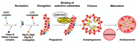
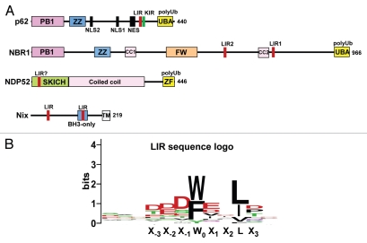
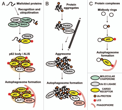
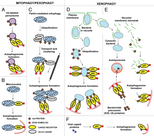
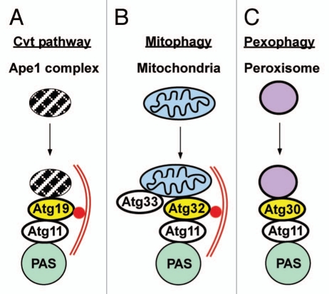
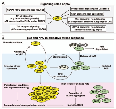
Mounting evidence suggests that autophagy is a more selective process than originally anticipated. The discovery and characterization of autophagic adapters, like p62 and NBR1, has provided mechanistic insight into this process. p62 and NBR1 are both selectively degraded by autophagy and able to act as cargo receptors for degradation of ubiquitinated substrates. A direct interaction between these autophagic adapters and the autophagosomal marker protein LC3, mediated by a so-called LIR (LC3-interacting region) motif, their inherent ability to polymerize or aggregate as well as their ability to specifically recognize substrates are required for efficient selective autophagy. These three required features of autophagic cargo receptors are evolutionarily conserved and also employed in the yeast cytoplasm-to-vacuole targeting (Cvt) pathway and in the degradation of P granules in C. elegans. Here, we review the mechanistic basis of selective autophagy in mammalian cells discussing the degradation of misfolded proteins, p62 bodies, aggresomes, mitochondria and invading bacteria. The emerging picture of selective autophagy affecting the regulation of cell signaling with consequences for oxidative stress responses, tumorigenesis and innate immunity is also addressed.


Figures

Figure 1
Figure 1
Model for selective autophagy in mammalian cells. Autophagosome formation is initiated (nucleation step) by the ULK1 complex and the class III PtdIns 3-kinase complex located at the phagophore. Additional ATG proteins are required for growth of the phagophore (elongation step), that depends on two Ub-like conjugation reactions. First, conjugation of ATG12 to ATG5 results in the formation of an oligomeric complex between the ATG12-ATG5 conjugate and ATG16L. This complex then acts as an E3 ligase assisting the E2 ATG3 in the lipidation of ATG8 family proteins at the phagophore. Selective autophagy depends on binding of substrates to the inner surface of the growing phagophore, and this can be achieved by cargo receptors that are associated both with the substrate and with lipidated ATG8 family proteins anchored to the phagophore. Aggregation of the substrate and/or cargo receptor is required for efficient sequestration. Closure results in the formation of a double-membrane autophagosome. Fusion of autophagosomes with late endosomes or lysosomes (maturation step) is then required for the formation of autolysomes where the substrates are degraded.
Figure 2
Figure 2
(A) Domain architecture of the hitherto characterized mammalian autophagic cargo receptors p62, NBR1, NDP52 and Nix. The FW domain has been named NBR1 box in a review by Kraft et al. (B) A consensus sequence logo for the LIR motif. The logo is based on 25 different LIR motifs from 21 different proteins that all bind directly to ATG8 family proteins.
Figure 3
Figure 3
Cargo receptors involved in degradation of proteins by selective autophagy. (A) Degradation of misfolded proteins via the formation of p62 bodies. Misfolded proteins are first recognized by molecular chaperones. Recognition by a complex of Bag3, HspB8 and Hsc70 and subsequent ubiquitination by an associated E3 ligase such as CHIP, results in the recruitment of p62. Together with its interaction partners NBR1 and ALFY, p62 then mediates the assembly of Ub-labeled misfolded proteins into p62 bodies of variable sizes. The contents of p62 bodies can be degraded by the proteasome or by autophagy and autophagic degradation depends on interactions of p62 and NBR1 with LC3, and ALFY with ATG5. (B) Degradation via formation of aggresomes. Aggresomes are formed by HDAC6-mediated transport of smaller Ub-containing aggregates to the microtubule-organizing center region. Recruitment of p62 and ALFY to the aggresomes may induce their degradation by autophagy. However, it is often unclear if it is the large aggregates or smaller precursors that are most efficiently degraded. (C) Degradation of protein complexes. Midbody rings are selectively degraded by autophagy after mitosis, and the process depends on their ubiquitination and on recruitment of p62.
Figure 4
Figure 4
Cargo receptors involved in mitophagy, pexophagy and xenophagy in mammalian cells. (A) Peroxisomes artificially labeled with monoUb on their surface are specifically recognized by p62 and degraded by autophagy, illustrating that Ub is a signal for p62-mediated autophagic degradation. (B) Nix-dependent mitophagy during development. Nix interacts with ATG8 family proteins, and acts as a cargo receptor for the delivery of mitochondria to the phagophore. The process is Ub-independent. (C) Removal of depolarized mitochondria by Parkin-mediated autophagy. Recruitment of Parkin to the mitochondria is regulated by PINK1. Parkin-mediated ubiquitination is essential and it recruits p62 and HDAC6. These proteins are responsible for a transport of depolarized mitochondria to the perinuclear region and for their assembly into clusters. It is not clear what cargo receptors are directly involved in the delivery of mitochondria to the phagophore. (D) Removal of intracellular bacteria by selective autophagy. Many intracellular bacteria such as S. enterica can reside and replicate within vacuolar structures, but they also leak out in the cytosol and are then recognized and ubiquitinated by an unknown machinery. Both p62 and NDP52 are recruited to ubiquitinated bacteria resulting in their delivery to the phagophore. (E) Some specialized pathogens have a cytosolic lifestyle. These bacteria may have developed specific systems to avoid ubiquitination, and fewer bacteria are then delivered to the autophagic system. p62 acts as a cargo receptor for the delivery of bactericidal precursors to the autolysosomes via selective autophagy, and for some pathogens such as M. tuberculosis, this may be essential for the killing of the bacteria. p62 also acts as a cargo receptor for autophagy of vacuolar membrane remnants after a bacterium escapes from the phagosome after cell entry.
Figure 5
Figure 5
Selective autophagy in yeast. (A) Atg19 is acting as a cargo receptor in the Cvt pathway. It binds to the Ape1 complex, and delivery to the phagophore is mediated by interactions with Atg11 located at the phagophore assembly site (PAS), and Atg8 attached to the phagophore membrane. (B) Atg32 is the cargo receptor in mitophagy. Atg32 is a mitochondrial outer membrane protein, and it interacts with Atg11 and Atg8 to mediate the delivery of mitochondria to the phagophore. (C) Atg30 is the cargo receptor in pexophagy. It is a peroxisomal membrane protein and interacts with PpAtg11, but does not interact with PpAtg8.
Figure 6
Figure 6
Signaling roles of p62 involving selective autophagy. (A) Signaling roles of p62 that may, at least in part, be regulated by autophagy. (B) p62 and Nrf2 as regulators of the oxidative stress response. Under normal conditions, there is a low level of p62 and Nrf2 due to selective autophagy of p62 and KEAP1-mediated proteasomal degradation of Nrf2. Under oxidative stress conditions, there is an elevated level of p62 and Nrf2. This results in the establishment of a feedback loop where Nrf2 induces expression of p62, and p62 inhibits KEAP1-mediated degradation of Nrf2. The net effect is induction of the intracellular antioxidant response. Under pathological conditions associated with inhibition of autophagy, there is a constitutive high level of p62 and Nrf2. This potentially induces ROS production, inhibits proteasomes and acts as a tumor-promoting factor.

References

    1. Levine B, Kroemer G. Autophagy in the pathogenesis of disease. Cell. 2008;132:27–42. - PMC - PubMed
    1. Xie Z, Klionsky DJ. Autophagosome formation: core machinery and adaptations. Nat Cell Biol. 2007;9:1102–1109. - PubMed
    1. Mizushima N. Autophagy: process and function. Genes Dev. 2007;21:2861–2873. - PubMed
    1. Deretic V. Autophagy in infection. Curr Opin Cell Biol. 2010;22:252–262. - PMC - PubMed
    1. Mizushima N, Levine B, Cuervo AM, Klionsky DJ. Autophagy fights disease through cellular self-digestion. Nature. 2008;451:1069–1075. - PMC - PubMed

Publication types

Substances

LinkOut - more resources