Clinical Trial
. 2011 May;19(5):990-9.
doi: 10.1038/mt.2010.289. Epub 2010 Dec 28.

Intradermal vaccinations with RNA coding for TAA generate CD8+ and CD4+ immune responses and induce clinical benefit in vaccinated patients

Affiliations
Clinical Trial

Intradermal vaccinations with RNA coding for TAA generate CD8+ and CD4+ immune responses and induce clinical benefit in vaccinated patients

Susanne M Rittig et al. Mol Ther. 2011 May.

Abstract

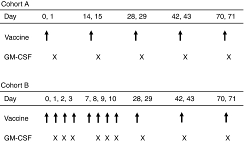
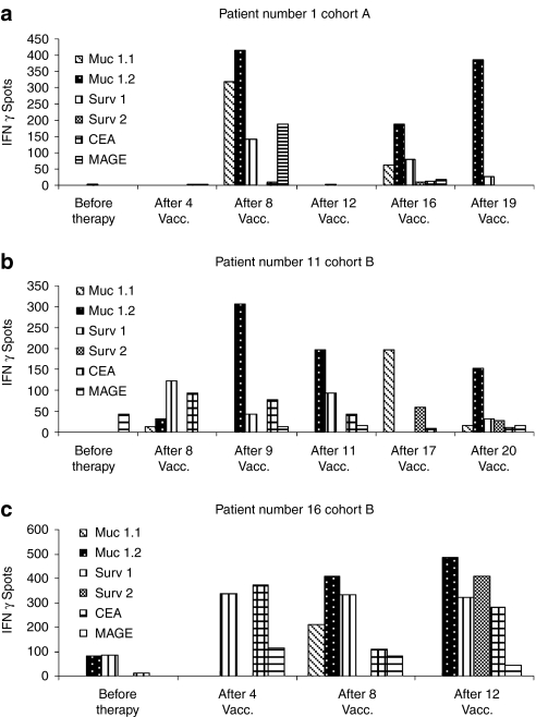
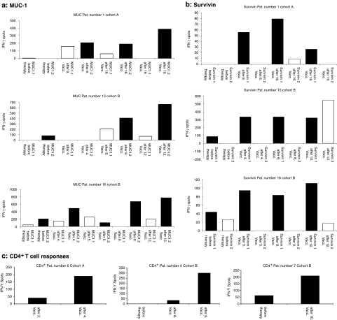
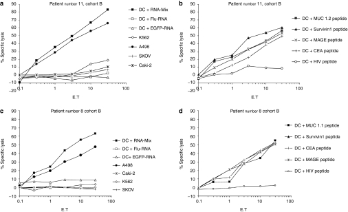
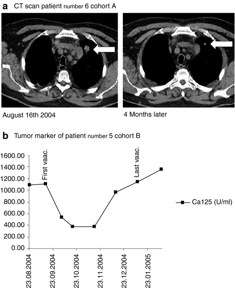
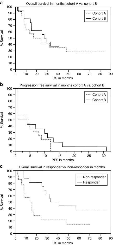
The aim of this phase I/II nonrandomized trial was to assess feasibility, safety as well as immunological and clinical responses of a mRNA-based vaccination in patients with stage IV renal cell cancer using granulocyte-macrophage colony stimulating factor (GM-CSF) as adjuvant. Intradermal injections of in vitro transcribed naked mRNA, which was generated using plasmids coding for the tumor-associated antigens mucin 1(MUC1), carcinoembryonic (CEA), human epidermal growth factor receptor 2 (Her-2/neu), telomerase, survivin, and melanoma-associated antigen 1 (MAGE-A1) were performed in 30 enrolled patients. In the first 14 patients (cohort A) vaccinations were administered on days 0, 14, 28, and 42 (20 µg/antigen) while in the consecutive 16 patients (cohort B) an intensified protocol consisting of injections at days 0-3, 7-10, 28, and 42 (50 µg/antigen) was used. In both cohorts, after this induction period, vaccinations were repeated monthly until tumor progression analyzed by Response Evaluation Criteria In Solid Tumors criteria (RECIST). Vaccinations were well tolerated with no severe side effects and induced clinical responses [six stable diseases (SD) and one partial response in cohort A and nine SD in cohort B]. In cohort A, 35.7% survived 4 years (median survival 24 months) compared to 31.25% in cohort B (median survival 29 months). Induction of CD4(+) and CD8(+) T cell responses was shown for several tumor-associated antigens (TAA) using interferon-γ (IFN-γ) enzyme-linked immunosorbent spot (ELISpot) and Cr-release assays.


Figures

Figure 1
Figure 1
Study design (injection schedule). Vaccinations were performed on days 0, 14, 28, and 42 in cohort A and on days 0–3, 7–10, 28, and 42 in cohort B (marked by the arrow). Vaccinations were repeated monthly until tumor progression. On the day following mRNA-injection, granulocyte-macrophage colony stimulating factor (GM-CSF) was applied subcutaneously (marked by “x”).
Figure 2
Figure 2
Generation of immunological responses upon vaccination with in vitro transcribed mRNA; exemplary interferon-γ (IFN-γ) enzyme-linked immunosorbent spot (ELISpot) assays: course of several tumor-associated antigen (TAA). Autologous peripheral blood mononuclear cells from human leukocyte antigen (HLA)-A2+ patients were pulsed with the peptides MUC 1.1, MUC 1.2, MAGE, CEA, survivin 1 and 2 deduced from TAA used in the vaccine and used as stimulators. (a–c) Representative ELISpot assays of several different T cell response patterns of three patients. Samples from prevaccination and representative samples after one or more vaccinations were evaluated simultaneously. CEA, carcinoembryonic; MAGE, melanoma-associated antigen 1; MUC 1, mucin 1.
Figure 3
Figure 3
Generation of immunological responses upon vaccination with in vitro transcribed mRNA. Exemplary enzyme-linked immunosorbent spot (ELISpot) assays. (a,b) Immunodominant antigens. Of all used antigens, immune responses for the epitopes MUC 1.2 and survivin 1 were pronounced in ELISpot assays indicating that these epitopes are immunodominant. Detailed course of immune responses are displayed for the epitopes (a) MUC 1.1 and 1.2 (b) and survivin 1 and 2 in three patients. (c) Results obtained for CD4+ cells. Autologous CD4+ T cells were isolated by magnetic activated cell sorting technology and stimulated with dendritic cells electroporated with a mixture of RNA coding for tumor-associated antigen used in the vaccine. Enhanced green fluorescent protein (EGFP)-RNA served as negative control, counted spots in EGFP vials were subtracted. MUC 1, mucin 1.
Figure 4
Figure 4
Lytic activity of vaccine-induced T lymphocytes. Cytotoxic T lymphocytes obtained after in vitro restimulation efficiently lysed target cells [dendritic cells (DC)] pulsed with the antigenic peptides (MUC1, survivin, MAGE, CEA), as well as DC electroporated with a mixture of RNA coding for tumor-associated antigen (TAA). Lysis was shown to be antigen-specific since no lysis was detected when target cells were loaded with the peptide derived from HIV or when electroporated with enhanced green fluorescent protein (EGFP) RNA. In addition, the human leukocyte antigen (HLA)-A2 positive tumor cell line A498 [renal cell carcinoma (RCC)], as well as HLA-A2 negative control cell lines like SK-OV-3 cells (ovarian cancer) and CaKi-2 (RCC) were included demonstrating HLA restricted lysis of CTL. K562 [chronic myelogenous leukemia (CML) in blast crisis] was used to exclude natural killer-cell mediated lysis. Data from two patients are presented (a,b: patient number 11 and c,d: patient number 8, cohort B). CEA, carcinoembryonic; MAGE, melanoma-associated antigen 1; MUC 1, mucin 1.
Figure 5
Figure 5
Clinical responses in vaccinated patients. (a) Computed tomography (CT) scan of patient number 6 (cohort A) with pulmonary metastases. Size of metatases diminished after 4 months of vaccination. (b) Course of tumor markers under vaccination (patient number 5, cohort B) The arrow marks the first and the last injection. The need of paracentesis diminished during vaccination.
Figure 6
Figure 6
Kaplan–Meier graphs: survival data. (a) Overall survival in months of patients in cohort A versus cohort B. Log-rank test showing no significant differences in overall survival (P = 0.85). Median survival cohort A: 24 months, cohort B: 29 months. (b) Progression free survival in months of patients in cohort A versus cohort B. Log rank test showing no significant differences in progression free survival (P = 0.33). (c) Overall survival in clinical responders versus nonresponders. Patients who had shown objective response or at least stable disease at the first staging time point were defined as clinical responders (P = 0.02). Median survival nonresponder: 14 months, responder: 41 months. PFS, progression free survival; OS, overall survival.

References

    1. Motzer RJ, Bander NH., and, Nanus DM. Renal-cell carcinoma. N Engl J Med. 1996;335:865–875. - PubMed
    1. Belldegrun A, Muul LM., and, Rosenberg SA. Interleukin 2 expanded tumor-infiltrating lymphocytes in human renal cell cancer: isolation, characterization, and antitumor activity. Cancer Res. 1988;48:206–214. - PubMed
    1. Thurnher M, Radmayr C, Ramoner R, Ebner S, Böck G, Klocker H, et al. Human renal-cell carcinoma tissue contains dendritic cells. Int J Cancer. 1996;68:1–7. - PubMed
    1. Michael A., and, Pandha HS. Renal-cell carcinoma: tumour markers, T-cell epitopes, and potential for new therapies. Lancet Oncol. 2003;4:215–223. - PubMed
    1. Renkvist N, Castelli C, Robbins PF., and, Parmiani G. A listing of human tumor antigens recognized by T cells. Cancer Immunol Immunother. 2001;50:3–15. - PMC - PubMed

Publication types

MeSH terms