

doi: 10.1038/nrmicro2475.
Epub 2010 Dec 29.
Genetic control of Candida albicans biofilm development
Affiliations
- PMID: 21189476
- PMCID: PMC3891587
- DOI: 10.1038/nrmicro2475
Item in Clipboard
Genetic control of Candida albicans biofilm development
Nat Rev Microbiol.
2011 Feb.
Abstract
Candida species cause frequent infections owing to their ability to form biofilms - surface-associated microbial communities - primarily on implanted medical devices. Increasingly, mechanistic studies have identified the gene products that participate directly in the development of Candida albicans biofilms, as well as the regulatory circuitry and networks that control their expression and activity. These studies have uncovered new mechanisms and signals that govern C. albicans biofilm development and associated drug resistance, thus providing biological insight and therapeutic foresight.
Figures
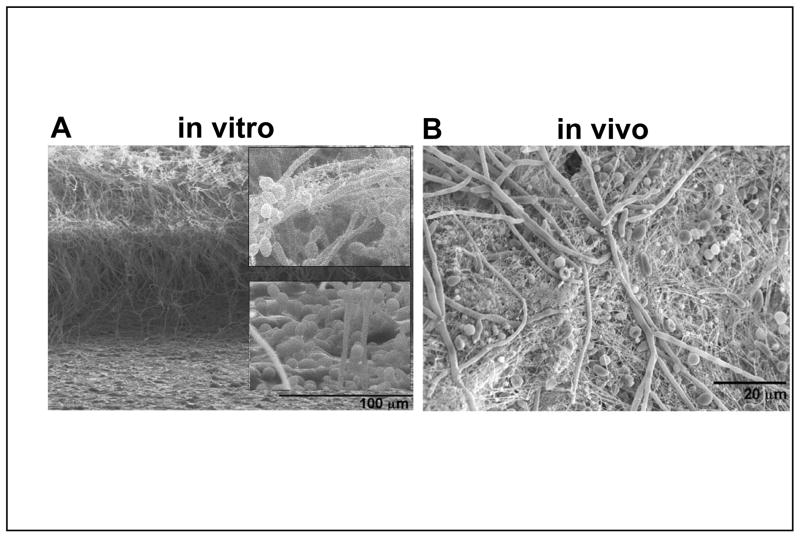
A) Scanning electron micrograph (SEM) of an in vitro biofilm. The biofilm sample was sliced to reveal three layers in a cross-sectional view (J. S. Finkel, J. Suhan, and A. P. Mitchell, unpublished results). The basal layer includes primarily yeast cells, as evident in the lower enlarged inset. The central layer is mainly hyphae. The upper layer has yeast cells budding from the hyphae. The upper enlarged inset shows extracellular matrix material, which appears fibrous in this preparation. B) SEM of an in vivo biofilm from the rat catheter model . Yeast cells, hyphae, and some pseudohyphal cells are evident, along with extracellular matrix material. (This image was provided by J. Nett and D. Andes.)
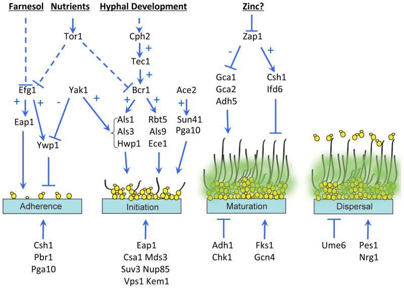
In the adherence step, yeast-form cells adhere to the substrate. In the initiation step, the cells propagate to form microcolonies and germ tubes form to yield hyphae. In the maturation step, the biofilm biomass expands, the extracellular matrix accumulates and drug resistance increases. In the dispersal step, yeast-form cells are released to colonize the surrounding environment. The upper half of the diagram depicts several known pathway relationships. The bottom half includes additional genes that function in a specific step, but may not be connected to a known pathway. For simplicity, some known pathway relationships have been omitted. A few genes are presented more than once if they have roles in more then one step of biofilm formation. Arrows represent positive relationships; T-shaped bars represent negative relationships. A “+” indicates that the upstream gene/signal stimulates expression of the downstream target; a “−” indicates that the upstream gene/signal inhibits expression of the downstream target. Dashed lines indicate repression by an indirect mechanism.
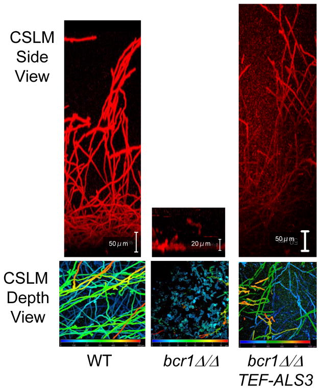
These panels are confocal scanning laser micrographs of concanavalin A-alexafluor stained biofilms, grown under standard in vitro conditions . The top panels are side views; the bottom panels are pseudocolor depth views, in which blue color represents cells closest to the substrate and red color represents cells farthest from the substrate. The wild type biofilm has a dense mixture of yeast cells and hyphae, which gradually becomes predominantly hyphae at the top of the biofilm. The bcr1Δ/Δ biofilm forms a basal layer of yeast cells attached to the substrate with little to no hyphae. Increased expression of ALS3 in the bcr1Δ/Δ strain permits substantial biofilm formation.
References
-
- Pfaller MA, Diekema DJ. Epidemiology of invasive mycoses in North America. Crit Rev Microbiol. 36:1–53. - PubMed
-
- Pappas PG, et al. Guidelines for treatment of candidiasis. Clin Infect Dis. 2004;38:161–89. - PubMed
-
- Costerton JW, Stewart PS, Greenberg EP. Bacterial biofilms: a common cause of persistent infections. Science. 1999;284:1318–22. - PubMed
-
- Douglas LJ. Candida biofilms and their role in infection. Trends Microbiol. 2003;11:30–6. - PubMed
MeSH terms
Grants and funding
LinkOut - more resources
Full Text Sources
Other Literature Sources
