Renal fibrosis
- PMID: 21189948
- PMCID: PMC3004484
- DOI: 10.3345/kjp.2010.53.7.735
Renal fibrosis
Abstract
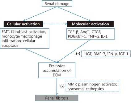
Renal fibrosis, characterized by tubulointerstitial fibrosis and glomerulosclerosis, is the final manifestation of chronic kidney disease. Renal fibrosis is characterized by an excessive accumulation and deposition of extracellular matrix components. This pathologic result usually originates from both underlying complicated cellular activities such as epithelial-to-mesenchymal transition, fibroblast activation, monocyte/macrophage infiltration, and cellular apoptosis and the activation of signaling molecules such as transforming growth factor beta and angiotensin II. However, because the pathogenesis of renal fibrosis is extremely complicated and our knowledge regarding this condition is still limited, further studies are needed.
Keywords: Fibrosis; Glomerulosclerosis; Kidney disease.
Figures

References
-
- Liu Y. Renal fibrosis: new insights into the pathogenesis and therapeutics. Kidney Int. 2006;69:213–217. - PubMed
-
- el Nahas AM, Muchaneta-Kubara EC, Essawy M, Soylemezoglu O. Renal fibrosis: insights into pathogenesis and treatment. Int J Biochem Cell Biol. 1997;29:55–62. - PubMed
-
- Eitner F, Floege J. Novel insights into renal fibrosis. Curr Opin Nephrol Hypertens. 2003;12:227–232. - PubMed
-
- Remuzzi G, Bertani T. Pathophysiology of progressive nephropathies. N Engl J Med. 1998;339:1448–1456. - PubMed
-
- Zeisberg M, Soubasakos MA, Kalluri R. Animal models of renal fibrosis. Methods Mol Med. 2005;117:261–272. - PubMed
LinkOut - more resources
Full Text Sources
Other Literature Sources