

Review
doi: 10.1007/s00431-010-1373-6.
Epub 2010 Dec 30.
Clinical practice: surgical approaches to urolithiasis in children
Affiliations
- PMID: 21190040
- PMCID: PMC4011548
- DOI: 10.1007/s00431-010-1373-6
Item in Clipboard
Review
Clinical practice: surgical approaches to urolithiasis in children
Eur J Pediatr.
2011 Jun.
Abstract
The incidence of urolithiasis in children is increasing. Adequate knowledge of treatment modalities and surgical options is therefore essential for every pediatrician. Surgical approaches to urolithiasis in children continue to evolve with advancements in technology and sophistication of current equipment and techniques. Perhaps the most significant development in new techniques is the advent of robotic-assisted laparoscopy. This review, for the general pediatrician, summarizes the most recent pediatric data and guidelines for surgical approaches to treatment of urolithiasis.
Conflict of interest statement
Figures
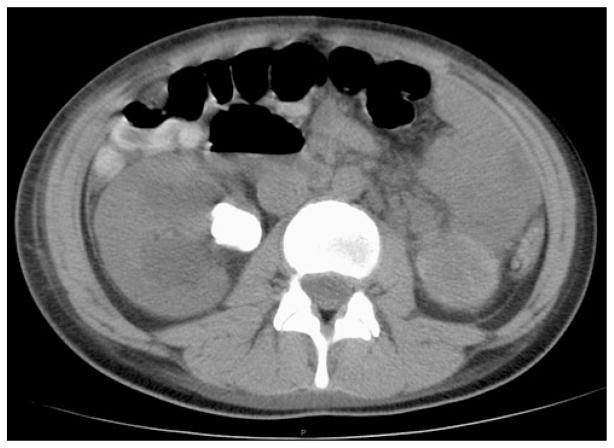
Computed tomography axial image of 2.5 cm right ureteropelvic junction stone causing hydronephrosis in 16-year-old female

Ultrasound image of a staghorn renal calculus in 6-year-old female. In this sagittally oriented image, multiple sections of this stone can be seen, appearing as hyperechoic areas with posterior shadowing. No dilation of the renal collecting system is present. Ultrasound can underestimate the size of the stone due to inadequate penetration through the stone of the sound waves
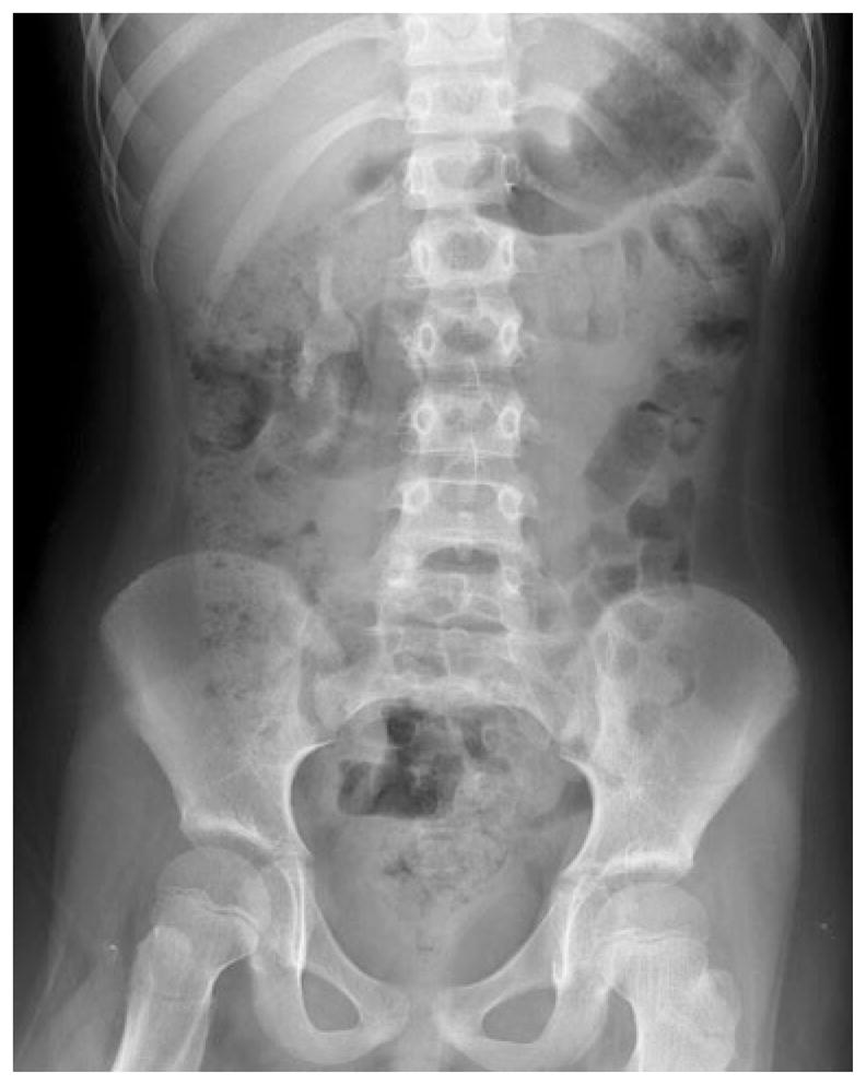
Plain film X-ray (KUB) of staghorn renal calculus shown in Fig. 2. Right-sided radio-opaque stone measures approximately 5×2 cm and fills the majority of the collecting system

Dornier HM3 lithotripter for extracorporeal lithotripsy. The patient under anesthesia is partially submerged in a water bath that facilitates energy transfer of the sound waves focused on the stone using fluoroscopic monitoring

Pediatric ESWL stone-free rates based on size of the studies and location of the stones. Clearance of lower pole stones is significantly less efficient than other locations. Adapted from Smaldone et al., [30]

Stone-free rates in children following PCNL and ESWL. Adapted from Smaldone et al., [30]
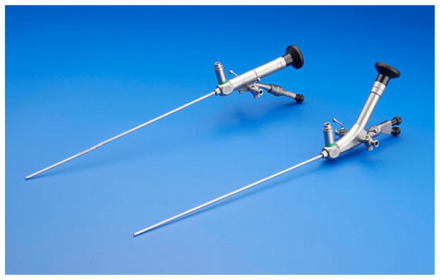
Rigid ureteroscopes with the smallest caliber at 4.5-Fr are useful for distal ureteral stones, but can reach into the proximal ureter safely. ©2009 Photo Courtesy of KARL STORZ Endoscopy-America, Inc

Flexible ureteroscope that is particularly useful for access to the kidney using a retrograde approach. ©2009 Photo Courtesy of KARL STORZ Endoscopy-America, Inc
References
-
- Ather MH, Noor MA. Does size and site matter for renal stones up to 30-mm in size in children treated by extracorporeal lithotripsy? Urology. 2003;61(1):212–215. S0090429502021283. discussion 215. - PubMed
-
- Badawy H, Salama A, Eissa M, et al. Percutaneous management of renal calculi: experience with percutaneous nephrolithotomy in 60 children. J Urol. 1999;162(5):1710–1713. S0022-5347(05)68220-1. - PubMed
Publication types
MeSH terms
Grants and funding
LinkOut - more resources
Full Text Sources
