Review
. 2011 Jun;68(11):1829-41.
doi: 10.1007/s00018-010-0611-4. Epub 2010 Dec 29.

Mining electron density for functionally relevant protein polysterism in crystal structures

Affiliations
Review

Mining electron density for functionally relevant protein polysterism in crystal structures

James S Fraser et al. Cell Mol Life Sci. 2011 Jun.

Abstract

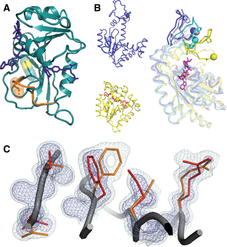
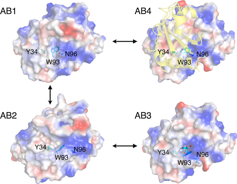
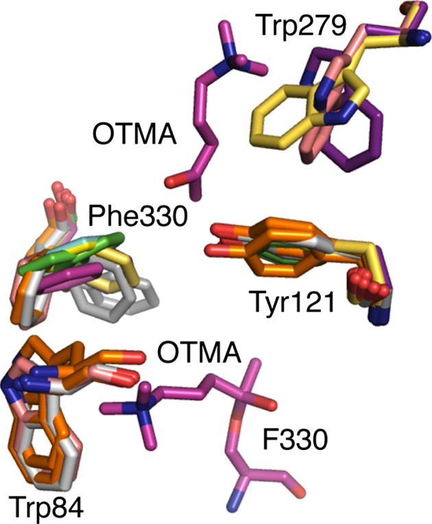
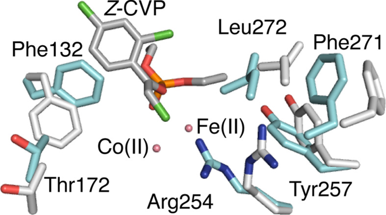
This review focuses on conceptual and methodological advances in our understanding and characterization of the conformational heterogeneity of proteins. Focusing on X-ray crystallography, we describe how polysterism, the interconversion of pre-existing conformational substates, has traditionally been analyzed by comparing independent crystal structures or multiple chains within a single crystal asymmetric unit. In contrast, recent studies have focused on mining electron density maps to reveal previously 'hidden' minor conformational substates. Functional tests of the importance of minor states suggest that evolutionary selection shapes the entire conformational landscape, including uniquely configured conformational substates, the relative distribution of these substates, and the speed at which the protein can interconvert between them. An increased focus on polysterism may shape the way protein structure and function is studied in the coming years.


Figures

Fig. 1
Fig. 1
Catalytically important motions detected by NMR relaxation experiments can arise through different structural mechanisms. a Polysterism revealed by comparison of NMR chemical shifts with reference to independent crystal structures in DHFR (1RC4 and 1RB2). The Met20 loop of DHFR samples a minor conformational substate (shown in orange) when bound to the substrates NADP+ and THF. The minor state chemical shift is correlated with the chemical shift of the major conformational substate for the immediately preceding state of the catalytic cycle. b Independent crystal structures of AdK in the apo/open state (blue 2RH5) and nucleotide-bound/closed state (yellow, with nucleotide in red 2RGX) reveal that subdomain lid closure underlies the NMR relaxation signal. In an orthogonal view, independent chains from one crystallographic asymmetric unit (blue, light blue, and cyan 2RH5) reveal that the transition path towards the closed state (yellow) is populated even when no nucleotide is bound. Residues on mobile loops are shown in stick and sphere to provide a visual landmark of the polysterism. c In the room-temperature structure of CypA (3K0N), interconverting side-chain conformations give rise to an NMR relaxation dispersion signal. The minor (orange) and major (red) conformational substates extending from the core (left) to the active site Arg55 (right) are shown within electron density mesh. Examining the electron density at low levels (light blue 0.3σ) below normal contour (dark blue 1σ) reveals the minor conformational substate
Fig. 2
Fig. 2
Hapten or protein binding selects pre-existing conformations of the antibody SPE7. Two distinct conformations of unbound SPE7 (AB1: 1OAQ; AB2: 1OCW) have been characterized in which part of the binding site consisting of Y34, W93 and N96 of the light chain are in different conformations. Owing to the orientation of W96, the binding site of the major conformation, AB1, is considerably ‘flatter’ than AB2. Ligand binding selects from these pre-existing substates, with protein-binding (AB4: 1OAZ) maintaining the AB1-like conformation of these residues and hapten-binding (DNP-Serine; AB3: 1OAU) maintaining an AB2-like conformation
Fig. 3
Fig. 3
Polysterism in the active site and peripheral site of acetylcholinesterase. The gateway to the active site from the peripheral anionic site in AChE is formed by F330 and Y121. The positioning of substrate in the active site and peripheral anionic site is shown by the substrate analog OTMA from the structure 2C5F. Several structures of AChE are overlayed showing the conformational flexibility of F330. These include: 2C4H (green), 2C58 (cyan), 2C5F (magenta), 2C5G (light yellow), 2V96 (light grey), 2V98 (orange), 1EA5 (pink), 2CEK (grey), 1ACJ (dark yellow). Polysterism in the ‘backdoor residue of the active site in shown by the structures 1EA5, 2V98, and 2V96. Polysterism of W279 at the peripheral anionic site is shown by 2CKM (purple), 1EA5 and 1ACJ
Fig. 4
Fig. 4
Major and minor conformations of the bacterial phosphotriesterase. The major ‘closed’ conformation of PTE is shown in cyan, while the minor ‘open’ conformation is shown in grey (3A4J). Both conformations were modeled into the electron density of an apo-enzyme crystal. The substrate Z-chlorfenvinphos is shown docked based on known enzyme:substrate complexes (2R1N). It can be seen that in the ‘closed’ conformation, several side-chains adopt rotamers that extend into the active site, greatly reducing its volume. One of these, F132, is shown to sterically clash with Z-chlorfenvinphos in its dominant conformation but to allow binding in its minor ‘open’ conformation
Fig. 5
Fig. 5
DNA binding allosterically modulates lever arm conformations and the transcriptional activation of glucocorticoid receptor. a The glucocorticoid receptor bound to the Sgk-sequence DNA (3G9P) adopts a dimeric structure with polysterism in the lever arm. b The electron density (dark blue 1σ, light blue 0.3σ) for the chain A (yellow) lever arm is well fit and suggests only small amplitude motion around the major conformation. c In contrast, despite originating from the same map, the electron density for the lever arm of chain B (black) for this area reveals potential polysterism. Although the lever arm is modeled into a different conformation, there is much unexplained density at low levels. d The conformations of the lever arms of chain A (yellow) and chain B (black) represent the major structure difference between the two chains. Low levels of electron density (light blue 0.3σ) shown from the area of chain B suggest that the conformation modeled for chain A may represent a component of the ensemble for both chains

References

    1. Hilser VJ, Garcia-Moreno EB, Oas TG, Kapp G, Whitten ST. A statistical thermodynamic model of the protein ensemble. Chem Rev. 2006;106:1545–1558. doi: 10.1021/cr040423+. - DOI - PubMed
    1. Kendrew JC, Dickerson RE, Strandberg BE, Hart RG, Davies DR, Phillips DC, Shore VC. Structure of myoglobin: a three-dimensional Fourier synthesis at 2 Å resolution. Nature. 1960;185:422–427. doi: 10.1038/185422a0. - DOI - PubMed
    1. Perutz MF, Rossmann MG, Cullis AF, Muirhead H, Will G, North AC. Structure of haemoglobin: a three-dimensional Fourier synthesis at 5.5-Å resolution, obtained by X-ray analysis. Nature. 1960;185:416–422. doi: 10.1038/185416a0. - DOI - PubMed
    1. Frauenfelder H, Sligar SG, Wolynes PG. The energy landscapes and motions of proteins. Science. 1991;254:1598–1603. doi: 10.1126/science.1749933. - DOI - PubMed
    1. Lang PT, Ng HL, Fraser JS, Corn JE, Echols N, Sales M, Holton JM, Alber T. Automated electron-density sampling reveals widespread conformational polymorphism in proteins. Protein Sci. 2010;19:1420–1431. doi: 10.1002/pro.423. - DOI - PMC - PubMed

Publication types