

Effect of the PARP-1 inhibitor PJ 34 on excitotoxic damage evoked by kainate on rat spinal cord organotypic slices
- PMID: 21190076
- PMCID: PMC11498577
- DOI: 10.1007/s10571-010-9640-7
Effect of the PARP-1 inhibitor PJ 34 on excitotoxic damage evoked by kainate on rat spinal cord organotypic slices
Abstract
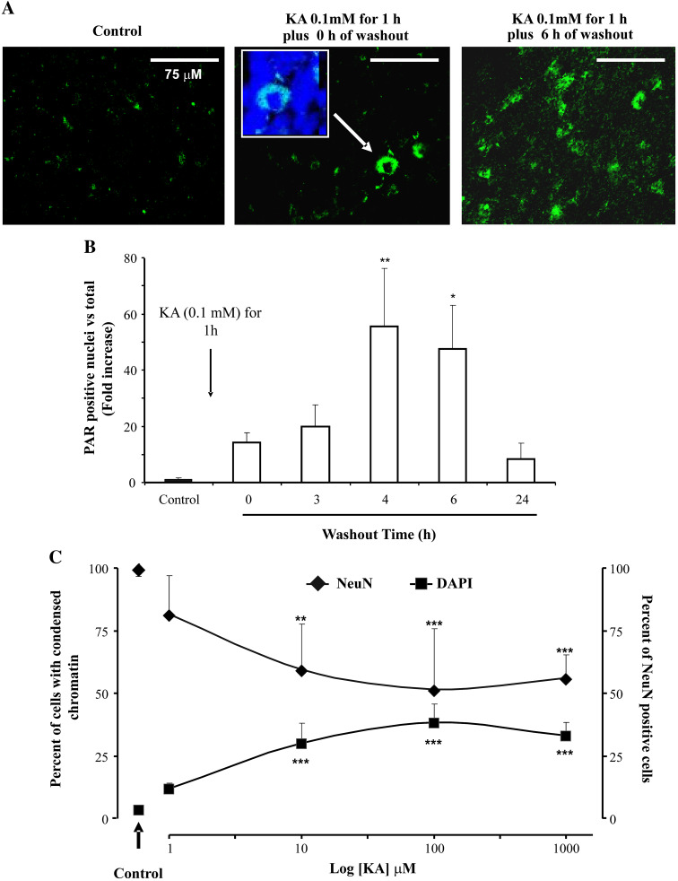
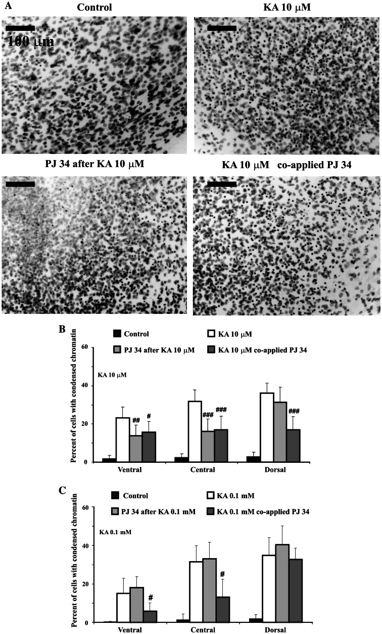
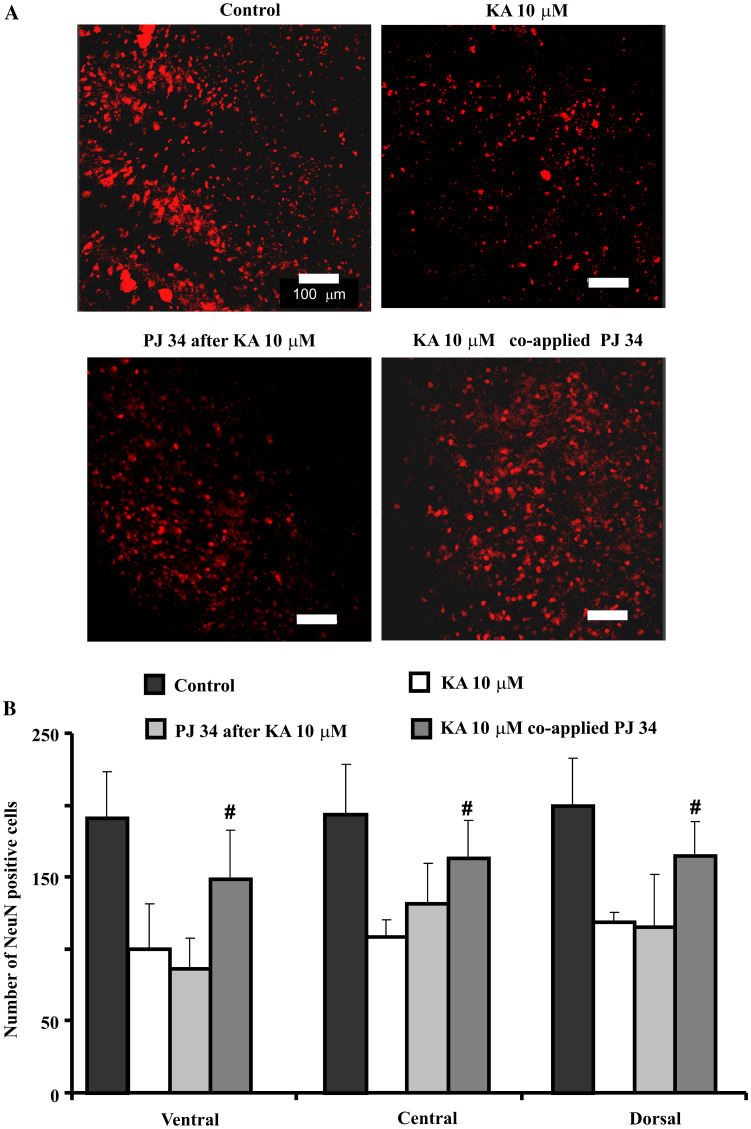
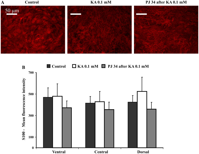
Excitotoxicity triggered by over-activation of glutamate receptors is thought to be an early mechanism of extensive neuronal death with consequent loss of function following lesion of spinal networks. One important process responsible for excitotoxic death is 'parthanatos' caused by hyperactivation of poly(ADP-ribose) polymerase (PARP) enzyme 1. Using rat organotypic spinal slices as in vitro models, the present study enquired if 2-(dimethylamino)-N-(5,6-dihydro-6-oxophenanthridin-2yl)acetamide (PJ 34), a pharmacological inhibitor of PARP-1, could counteract the excitotoxic damage evoked by transient application (1 h) of kainate, a potent analogue of glutamate. Kainate induced dose-dependent (1 μM threshold) neuronal loss (without damage to astrocytes) detected 24 h later via a PARP-1 dependent process that had peaked at 4 h after washout kainate. All spinal regions (ventral, central and dorsal) were affected, even though the largest damage was found in the dorsal area. Whereas PJ 34 did not protect against a large concentration (100 μM) of kainate, it significantly inhibited neuronal losses evoked by 10 μM kainate as long as it was co-applied with this glutamate agonist. When the application of PJ 34 was delayed to the washout time, neuroprotection was weak and regionally restricted. These data suggest that kainate-induced parthanatos developed early and was prevented by PJ 34 only when it was co-applied together with excitotoxic stimulus. Our results highlight the difficulty to arrest parthanatos as a mechanism of spinal neuron death in view of its low threshold of activation by kainate, its widespread distribution, and relatively fast development.
Figures
Similar articles
-
Studies of locomotor network neuroprotection by the selective poly(ADP-ribose) polymerase-1 inhibitor PJ-34 against excitotoxic injury to the rat spinal cord in vitro.Eur J Neurosci. 2011 Jun;33(12):2216-27. doi: 10.1111/j.1460-9568.2011.07714.x. Epub 2011 May 30. Eur J Neurosci. 2011. PMID: 21623955
-
Unusual increase in lumbar network excitability of the rat spinal cord evoked by the PARP-1 inhibitor PJ-34 through inhibition of glutamate uptake.Neuropharmacology. 2012 Sep;63(3):415-26. doi: 10.1016/j.neuropharm.2012.04.014. Epub 2012 Apr 28. Neuropharmacology. 2012. PMID: 22561282
-
Kainate-mediated excitotoxicity induces neuronal death in the rat spinal cord in vitro via a PARP-1 dependent cell death pathway (Parthanatos).Cell Mol Neurobiol. 2010 Oct;30(7):1001-12. doi: 10.1007/s10571-010-9531-y. Epub 2010 May 26. Cell Mol Neurobiol. 2010. PMID: 20502958 Free PMC article.
-
Poly(ADP-Ribose) polymerase-1 in acute neuronal death and inflammation: a strategy for neuroprotection.Ann N Y Acad Sci. 2003 May;993:217-28; discussion 287-8. doi: 10.1111/j.1749-6632.2003.tb07532.x. Ann N Y Acad Sci. 2003. PMID: 12853316 Review.
-
[Neuron damage in the rat spinal cord induced by acromelic acid].Rinsho Shinkeigaku. 1991 Dec;31(12):1313-5. Rinsho Shinkeigaku. 1991. PMID: 1817797 Review. Japanese.
Cited by
-
GABAergic Mechanisms Can Redress the Tilted Balance between Excitation and Inhibition in Damaged Spinal Networks.Mol Neurobiol. 2021 Aug;58(8):3769-3786. doi: 10.1007/s12035-021-02370-5. Epub 2021 Apr 7. Mol Neurobiol. 2021. PMID: 33826070 Free PMC article. Review.
-
PARP Inhibition Prevents Ethanol-Induced Neuroinflammatory Signaling and Neurodegeneration in Rat Adult-Age Brain Slice Cultures.J Pharmacol Exp Ther. 2018 Apr;365(1):117-126. doi: 10.1124/jpet.117.245290. Epub 2018 Jan 16. J Pharmacol Exp Ther. 2018. PMID: 29339456 Free PMC article.
-
PARP-1 inhibitors DPQ and PJ-34 negatively modulate proinflammatory commitment of human glioblastoma cells.Neurochem Res. 2013 Jan;38(1):50-8. doi: 10.1007/s11064-012-0887-x. Epub 2012 Sep 26. Neurochem Res. 2013. PMID: 23011206
-
Organotypic Spinal Cord Culture: a Proper Platform for the Functional Screening.Mol Neurobiol. 2016 Sep;53(7):4659-74. doi: 10.1007/s12035-015-9403-z. Epub 2015 Aug 27. Mol Neurobiol. 2016. PMID: 26310972 Review.
-
Excitotoxic cell death induces delayed proliferation of endogenous neuroprogenitor cells in organotypic slice cultures of the rat spinal cord.Cell Death Dis. 2013 Oct 31;4(10):e902. doi: 10.1038/cddis.2013.431. Cell Death Dis. 2013. PMID: 24176860 Free PMC article.
References
-
- Abdelkarim GE, Gertz K, Harms C, Katchanov J, Dirnagl U, Szabo C, Endres M (2001) Protective effects of PJ34, a novel, potent inhibitor of poly(ADP-ribose) polymerase (PARP) in in vitro and in vivo models of stroke. Int J Mol Med 7:255–260 - PubMed
-
- Agrawal SM, Lau L, Yong VW (2008) MMPs in the central nervous system: where the good guys go bad. Semin Cell Dev Biol 19:42–51 - PubMed
Publication types
MeSH terms
Substances
LinkOut - more resources
Full Text Sources
Miscellaneous
