

NMR relaxation and magnetic properties of superparamagnetic nanoworms
- PMID: 21190269
- PMCID: PMC6589091
- DOI: 10.1002/cmmi.387
NMR relaxation and magnetic properties of superparamagnetic nanoworms
Abstract
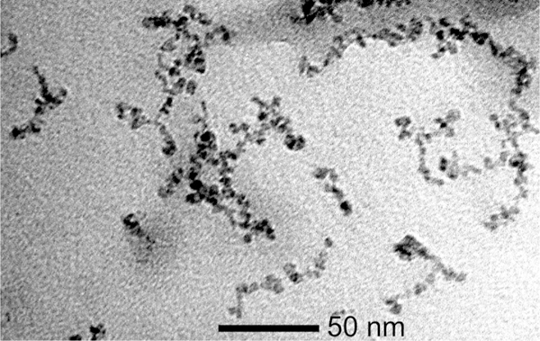
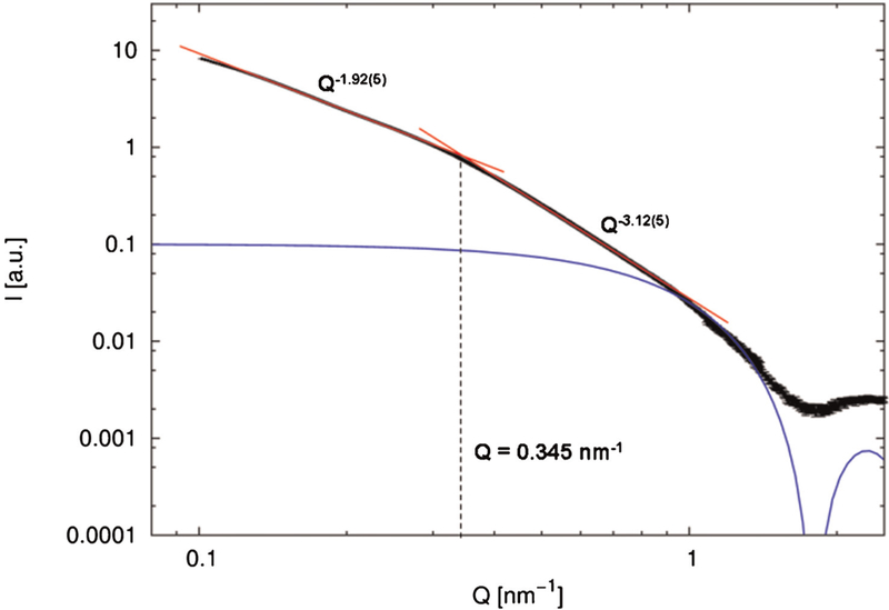
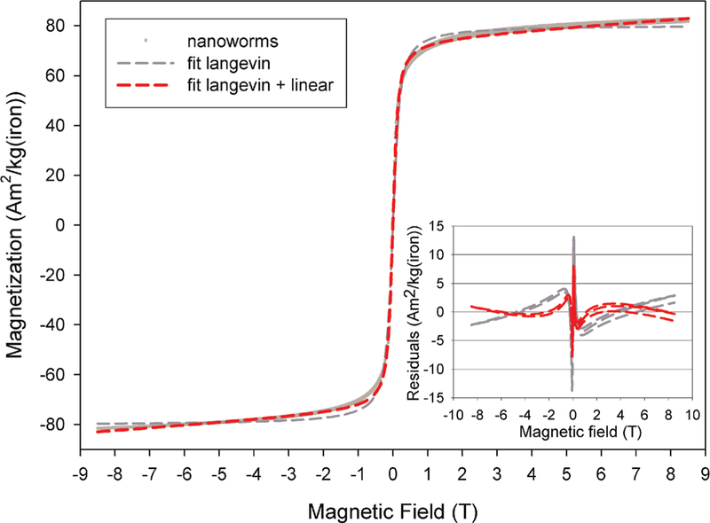
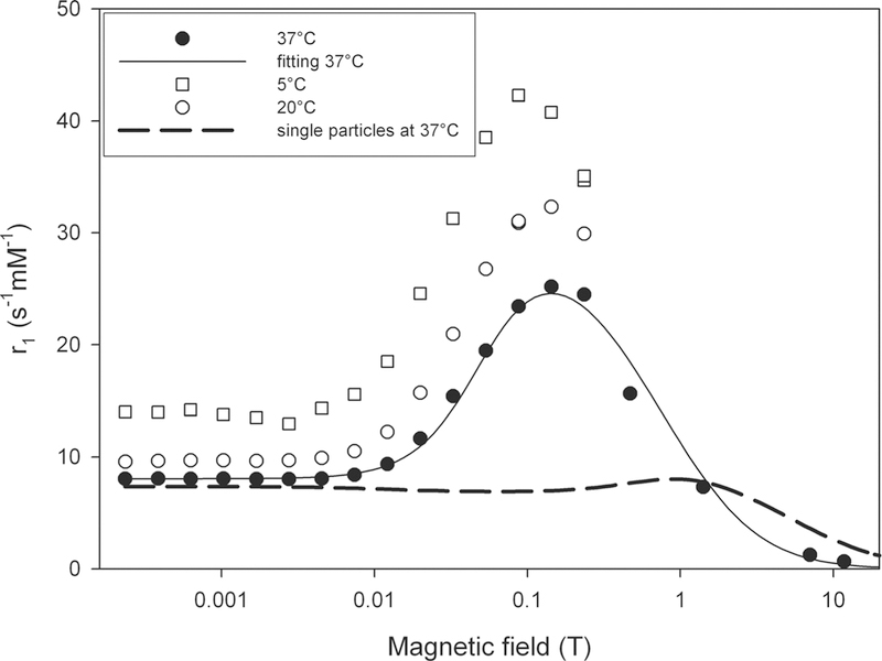
Maghemite particles are used as T₂ contrast agents for magnetic resonance imaging, especially for molecular and cellular imaging. Linear clusters of particles - called nanoworms - were recently developed to enhance the targeting efficiency. In this work, the magnetic and NMR relaxation properties of these nanoworms are studied at multiple magnetic fields. After the usual saturation at 0.5 T, the magnetization of the worms is still increasing, which results in an appreciable increase of the transverse relaxivity at high magnetic fields. The obtained relaxivities are typical of superparamagnetic particles of iron oxide (SPIOs). The transverse relaxation of the worms is clearly more efficient than for the isolated grains, which is confirmed by computer simulations. At high field, the longitudinal relaxation of the worms is less pronounced than for the grains, as expected for SPIOs. The nanoworms thus constitute a promising T₂ agent for cellular and molecular imaging.
Copyright © 2010 John Wiley & Sons, Ltd.
Figures
Similar articles
-
Magnetic resonance relaxation properties of superparamagnetic particles.Wiley Interdiscip Rev Nanomed Nanobiotechnol. 2009 May-Jun;1(3):299-310. doi: 10.1002/wnan.36. Wiley Interdiscip Rev Nanomed Nanobiotechnol. 2009. PMID: 20049798 Review.
-
A universal scaling law to predict the efficiency of magnetic nanoparticles as MRI T(2)-contrast agents.Adv Healthc Mater. 2012 Jul;1(4):502-12. doi: 10.1002/adhm.201200078. Epub 2012 May 16. Adv Healthc Mater. 2012. PMID: 23184784
-
Ferrimagnetic susceptibility contrast agents.Acta Radiol Suppl. 1993;387:1-30. Acta Radiol Suppl. 1993. PMID: 8390776
-
Increased transverse relaxivity in ultrasmall superparamagnetic iron oxide nanoparticles used as MRI contrast agent for biomedical imaging.Contrast Media Mol Imaging. 2016 Sep;11(5):350-361. doi: 10.1002/cmmi.1698. Epub 2016 May 27. Contrast Media Mol Imaging. 2016. PMID: 27230705
-
Proton relaxation enhancement.J Magn Reson Imaging. 1993 Jan-Feb;3(1):149-56. doi: 10.1002/jmri.1880030127. J Magn Reson Imaging. 1993. PMID: 8428082 Review.
Cited by
-
Comparing the signal enhancement of a gadolinium based and an iron-oxide based contrast agent in low-field MRI.PLoS One. 2021 Aug 17;16(8):e0256252. doi: 10.1371/journal.pone.0256252. eCollection 2021. PLoS One. 2021. PMID: 34403442 Free PMC article.
-
Stimuli-Responsive Iron Oxide Nanotheranostics: A Versatile and Powerful Approach for Cancer Therapy.Adv Healthc Mater. 2021 Mar;10(5):e2001044. doi: 10.1002/adhm.202001044. Epub 2020 Nov 23. Adv Healthc Mater. 2021. PMID: 33225633 Free PMC article. Review.
-
Imaging metastasis using an integrin-targeting chain-shaped nanoparticle.ACS Nano. 2012 Oct 23;6(10):8783-95. doi: 10.1021/nn303833p. Epub 2012 Sep 24. ACS Nano. 2012. PMID: 23005348 Free PMC article.
-
Assembly of linear nano-chains from iron oxide nanospheres with asymmetric surface chemistry.PLoS One. 2011 Jan 6;6(1):e15927. doi: 10.1371/journal.pone.0015927. PLoS One. 2011. PMID: 21253600 Free PMC article.
-
On-command drug release from nanochains inhibits growth of breast tumors.Pharm Res. 2014 Jun;31(6):1460-8. doi: 10.1007/s11095-013-1102-8. Epub 2013 Aug 9. Pharm Res. 2014. PMID: 23934254 Free PMC article.
References
-
- Bulte JW, Kraitchman DL. Iron oxide MR contrast agents for molecular and cellular imaging. NMR Biomed 2004; 17: 484–499. - PubMed
-
- Wang YX, Hussain SM, Krestin GP. Superparamagnetic iron oxide contrast agents:Physicochemical characteristics and applications in MR imaging. Eur Radiol 2001; 11: 2313–2319. - PubMed
-
- Gossuin Y, Gillis P, Hocq A, Vuong QL, Roch A. MR relaxation properties of superparamagnetic iron oxide particles. Nanomed Nanobiotechnol 2009; 1: 299–310. - PubMed
-
- Bulte JW, Vymazal J, Brooks RA, Pierpaoli C, Frank JA. Frequency dependence of MR relaxation times II. Iron oxides. J Magn Reson Imag 1993; 3: 641–648. - PubMed
-
- Bulte JWM, Brooks RA, Moskowitz BM, Bryant LH, Frank JA. Relaxometry and magnetometry of the MR contrast agent MION-46L. Magn Reson Med 1999; 42: 379–384. - PubMed
Publication types
MeSH terms
Substances
Grants and funding
LinkOut - more resources
Full Text Sources
Medical
