

Review
doi: 10.1056/NEJMra0808281.
General anesthesia, sleep, and coma
Affiliations
- PMID: 21190458
- PMCID: PMC3162622
- DOI: 10.1056/NEJMra0808281
Item in Clipboard
Review
General anesthesia, sleep, and coma
N Engl J Med.
.
No abstract available
Figures
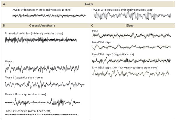
Panel A shows the EEG patterns when the patient is awake, with eyes open (left) and the alpha rhythm (10 Hz) with eyes closed (right). Panel B shows the EEG patterns during the states of general anesthesia: paradoxical excitation, phases 1 and 2, burst suppression, and the isoelectric tracing. Panel C shows the EEG patterns during the stages of sleep: rapid-eye-movement (REM) sleep; stage 1 non-REM sleep; stage 2 non-REM sleep, and stage 3 non-REM (slow-wave) sleep. The EEG patterns during recovery from coma — coma, vegetative state, and minimally conscious state — resemble the patterns during general anesthesia, sleep, and the awake state. EEG tracings during sleep are from Watson et al.
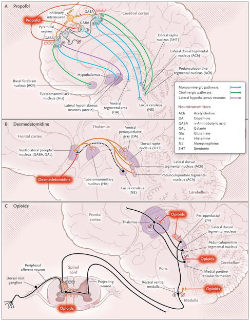
Panel A shows a GABAergic inhibitory interneuron (orange) synapsing on a pyramidal neuron (gray) receiving excitatory inputs from ascending arousal pathways. The monoaminergic pathways arise from the locus ceruleus, which releases norepinephrine; the raphe, which releases serotonin; the tuberomammillary nucleus, which releases histamine; and the ventral teg-mental area, which releases dopamine. The cholinergic pathways, which release acetylcholine, arise from the basal forebrain, the lateral dorsal tegmental nuclei, and the pedunculopontine tegmental nuclei. Lateral hypothalamic neurons release orexin. Propofol binds post-synaptically and enhances GABAergic inhibition, counteracting arousal inputs to the pyramidal neuron, decreasing its excitatory activity, and contributing to unconsciousness. Dexmedetomidine binds to α2 receptors on neurons from the locus ceruleus, inhibiting norepinephrine release (dashed line) in the ventrolateral preoptic nucleus, as shown in Panel B. The disinhibited ventrolateral preoptic nucleus reduces arousal by means of GABAA-mediated and galanin-mediated inhibition of the midbrain, hypothalamic, and pontine arousal nuclei. As shown in Panel C, opioids reduce arousal by inhibiting the release of acetylcholine from neurons projecting from the lateral dorsal and pedunculopontine tegmental nuclei to the medial pontine reticular formation and to the thalamus, by binding to opioid receptors in the periaqueductal gray and rostral ventral medulla, and by binding presynaptically and postsynaptically to spinal cord opioid receptors at the synapses between peripheral afferent neurons in the dorsal-root ganglion and projecting neurons.
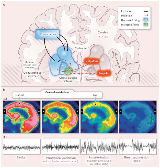
Cortical damage causes loss of excitatory inputs from the frontal cortex to the median spiny neurons in the striatum, as shown in Panel A. Normal striatal inhibition of the globus pallidus interna is lost, and the globus pallidus interna tonically inhibits the thalamus. Zolpidem and propofol may bind to GABAA1 receptors in the globus pallidus interna, blocking its inhibitory inputs to the thalamus; as a result, excitatory cortical inputs from the thalamus are restored, causing paradoxical excitation. Panel B schematically depicts changes in cerebral metabolism as measured by positron emission tomographic (PET) scanning and on electroencephalography (EEG) at different stages of coma recovery. In the awake state, the EEG pattern is active and cerebral metabolism is globally active. Paradoxical excitation induced by the administration of zolpidem, which is associated with behavioral improvement in some minimally conscious patients, is reflected by an active EEG pattern with reduced prefrontal cortex metabolism. Patients in minimally conscious and vegetative states may show EEG anteriorization, with alpha, theta, and delta EEG patterns and decreased metabolism in the frontal cortex, striatum, and thalamus. Burst suppression in coma correlates with globally depressed metabolism. General anesthesia results in similar EEG patterns. A denotes anterior, and P posterior.
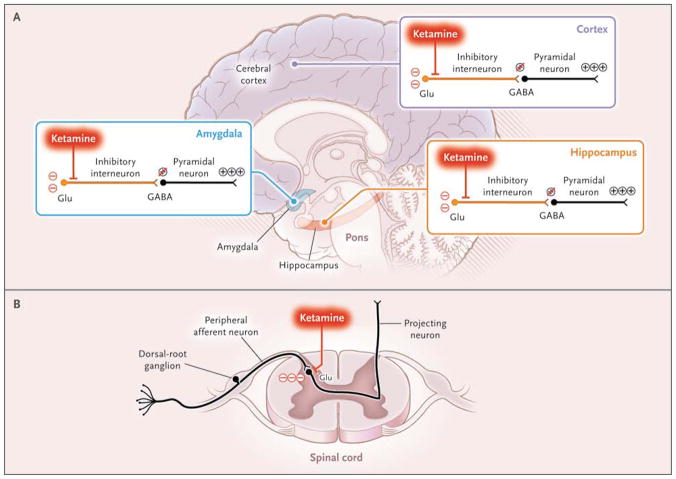
Ketamine binds preferentially to N-methyl-D-aspartate (NMDA) receptors on inhibitory interneurons in the cortex, limbic system (amygdala), and hippocampus, promoting an uncoordinated increase in neural activity, an active electroencephalographic pattern, and unconsciousness, as shown in Panel A. In the spinal cord, ketamine decreases arousal by blocking NMDA glutamate (Glu)–mediated nociceptive signals from peripheral afferent neurons in the dorsal-root ganglion to projecting neurons, as shown in Panel B.
References
-
- Sentinel event alert: preventing, and managing the impact of, anesthesia awareness. Oakbrook Terrace, IL: The Joint Commission; 2004. ( http://www.jointcommission.org/sentinel_event_alert_issue_32_preventing_....) - PubMed
-
- Evers A, Crowder M. Cellular and molecular mechanisms of anesthesia. In: Barash PG, Cullen BF, Stoelting RK, Cahalan M, Stock MC, editors. Clinical anesthesia. 6. New York: Lippincott Williams & Wilkins; 2006. pp. 95–114.
-
- Gibbs FA, Gibbs LE, Lennox WG. Effects on the electroencephalogram of certain drugs which influence nervous activity. Arch Intern Med. 1937;60:154–66.
-
- Kiersey DK, Bickford RG, Faulconer A., Jr Electro-encephalographic patterns produced by thiopental sodium during surgical operations; description and classification. Br J Anaesth. 1951;23:141–52. - PubMed
-
- Watson C, Bagdoyan H, Lydic R. A neurochemical perspective on states of consciousness. In: Hudetz AG, Pearce RA, editors. Suppressing the mind: anesthetic modulation of memory and consciousness. New York: Springer/Humana Press; 2010. pp. 33–80.
Publication types
MeSH terms
Grants and funding
LinkOut - more resources
Full Text Sources
Other Literature Sources
Medical