. 2011 Jan 5;100(1):183-92.
doi: 10.1016/j.bpj.2010.11.037.

Stoichiometry and affinity of the human serum albumin-Alzheimer's Aβ peptide interactions

Affiliations

Stoichiometry and affinity of the human serum albumin-Alzheimer's Aβ peptide interactions

Julijana Milojevic et al. Biophys J. .

Abstract

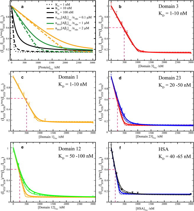
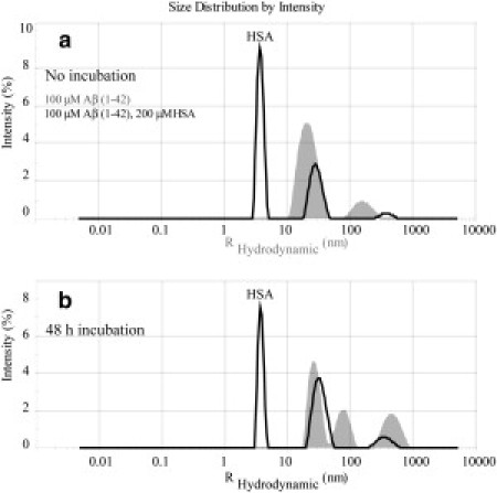
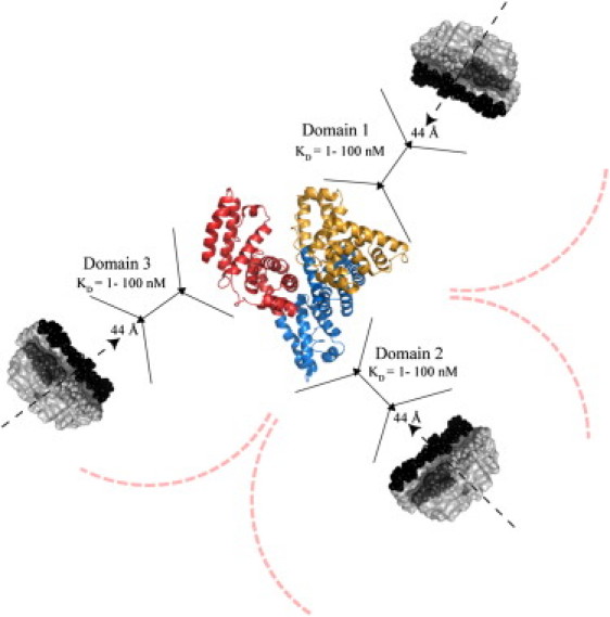
A promising strategy to control the aggregation of the Alzheimer's Aβ peptide in the brain is the clearance of Aβ from the central nervous system into the peripheral blood plasma. Among plasma proteins, human serum albumin plays a critical role in the Aβ clearance to the peripheral sink by binding to Aβ oligomers and preventing further growth into fibrils. However, the stoichiometry and the affinities of the albumin-Aβ oligomer interactions are still to be fully characterized. For this purpose, here we investigate the Aβ oligomer-albumin complexes through a novel and generally applicable experimental strategy combining saturation transfer and off-resonance relaxation NMR experiments with ultrafiltration, domain deletions, and dynamic light scattering. Our results show that the Aβ oligomers are recognized by albumin through sites that are evenly partitioned across the three albumin domains and that bind the Aβ oligomers with similar dissociation constants in the 1-100 nM range, as assessed based on a Scatchard-like model of the albumin inhibition isotherms. Our data not only explain why albumin is able to inhibit amyloid formation at physiological nM Aβ concentrations, but are also consistent with the presence of a single high affinity albumin-binding site per Aβ protofibril, which avoids the formation of extended insoluble aggregates.


Figures

Figure 1
Figure 1
Domain organization of HSA and design of the HSA constructs used in this investigation. (a) Ribbon diagram of the HSA structure in the fatty acid bound state (PDB file1E7H) (11). Palmitate molecules are shown in space-filling representation to indicate fatty acid binding sites. Domain 1 contains FA1 and FA2 binding sites, while domain 2 contains FA6 and FA7 binding sites. Domain 3 contains FA 3-5 binding sites. Drug binding sites are indicated as Sudlow sites 1 and 2, and their representative drug ligands are listed (10). (b) Constructs used in this study.
Figure 2
Figure 2
Effect of wt HSA and its deletion mutants on the one-dimensional NMR spectra of the Aβ (12–28) peptide. (a) Spectrum of 30 kDa filtered 1 mM Aβ (12–28). (b) Addition of 25 mM NaCl causes significant aggregation as indicated by line broadening and intensity losses. (cg) Effect of the addition of HSA and its deletion mutants: domains 12 and 23 and domains 3 and 1, respectively. A similar line sharpening is obtained upon addition of all protein constructs. (Dotted lines and arrows) Comparison of the one-dimensional intensities between different spectra; although protein addition results in line sharpening, it does not result in the restoration of the starting signal intensity. Spectra ac were previously published elsewhere (4) and are reported here only for comparison purposes.
Figure 3
Figure 3
Dose-response STD-based profiles for the inhibition of the Aβ (12–28) self-association by HSA and its deletion mutants. (a) Effect of different KD values and of different HSA-binding competent Aβ oligomer concentrations on dose-response STD curves simulated according to a Scatchard-like model. This model assumes full equivalence and independence of all sites in HSA that can bind Aβ oligomers. Further details on this model are available in the text. (bf) Effect of HSA deletion mutants (i.e., domain 3, domain 1, domains 23 and 13) and wt HSA, respectively, on the relative ISTD/ISTR ratios measured for the filtered Aβ (12–28) peptide aggregated through the addition of 25 mM NaCl. All ratios were normalized to their maximum value measured before protein addition. (b and c, Dashed and solid lines) Backcalculated dose-response curves using the Scatchard-like model and KD values of 1 and 10 nM, respectively. (d, Dashed and solid blue lines) Backcalculated dose-response curves using the Scatchard-like model and KD values of 20 and 50 nM, respectively. This range of KD values is in good agreement with the experimental data measured for the domain 23 construct. (Red lines) Obtained based on the single domain constructs KD values (1 and 10 nM), assuming that individual domains bind independently, i.e., simply downscaling the one-domain curves of panel b by a factor of two. (e, f, green and black curves) Computed using the KD values that fit the experimental data. (e, f, orange and blue lines) Obtained based on the one-domain curves of panel c, assuming fully independent binding of Aβ oligomers to two and three domains, i.e., simply downscaling the curves of panel c by a factor of two or three, respectively.
Figure 4
Figure 4
Correlations between the nonselective off-resonance relaxation data of full-length HSA and those measured for the four HSA deletion mutants shown in Fig. 1b. (Horizontal axes) Differences in residue-specific Hα nonselective off-resonance relaxation rates measured for the aggregated 1 mM Aβ (12–28) sample before and after addition of 10 μM full-length HSA. (ad, Vertical axes) Variations caused by the addition to the aggregated sample of 10 μM of the four HSA deletion mutants (domain 1, domain 3, and domains 12 and 23, respectively).
Figure 5
Figure 5
Intensity versus size distribution obtained from DLS measurements of 0.1 mM Aβ (1–42) in the presence and absence of 200 μM of HSA. (a) Measurements collected immediately after samples were prepared. (b) Measurements on the same samples incubated for 48 h at 37°C after preparation. (Shaded and solid representations) DLS profiles in the absence and presence of HSA, respectively. Note that all measurements were performed using 12-μL volume cells and a Zetasizer Nano S System (Malvern Instruments, Malvern, Worcestershire, UK) at 25°C.
Figure 6
Figure 6
Schematic model to summarize the prevailing stoichiometries and affinities for the complexes between HSA and the Aβ protofibrils. The structures of apo albumin (PDB file 1AO6) and of the Aβ protofibrils (PDB file 2BEG) were used to generate a model with reliable relative scales. The curved dashed lines indicate possible steric hindrance between Aβ protofibrils binding to different domains. The black solid lines are used as an aid in the comparison of the sizes of the albumin domains and of Aβ protofibril. The dashed arrows indicate the direction of protofibril growth while 44 Å corresponds to the protofibrils width. The C-terminal hydrophobic residues are depicted in black, whereas hydrophobic residues in the central hydrophobic core (CHC) are indicated in dark gray. Although a single Aβ protofibril is shown per albumin domain, we cannot rule out based on our data the presence of additional Aβ protofibrils.

References

    1. Deane R., Wu Z.H., Zlokovic B.V. RAGE (yin) versus LRP (yang) balance regulates Alzheimer amyloid β-peptide clearance through transport across the blood-brain barrier. Stroke. 2004;35(Suppl 1):2628–2631. - PubMed
    1. Llewellyn D.J., Langa K.M., Lang I.A. Serum albumin concentration and cognitive impairment. Curr. Alzheimer Res. 2010;7:91–96. - PMC - PubMed
    1. Boada M., Ortiz P., Páez A. Amyloid-targeted therapeutics in Alzheimer's disease: use of human albumin in plasma exchange as a novel approach for Aβ mobilization. Drug News Perspect. 2009;22:325–339. - PubMed
    1. Milojevic J., Raditsis A., Melacini G. Human serum albumin inhibits Aβ fibrilization through a “monomer-competitor” mechanism. Biophys. J. 2009;97:2585–2594. - PMC - PubMed
    1. Milojevic J., Esposito V., Melacini G. Understanding the molecular basis for the inhibition of the Alzheimer's Aβ-peptide oligomerization by human serum albumin using saturation transfer difference and off-resonance relaxation NMR spectroscopy. J. Am. Chem. Soc. 2007;129:4282–4290. - PubMed

Publication types