

X-ray diffraction from membrane protein nanocrystals
- PMID: 21190672
- PMCID: PMC3010013
- DOI: 10.1016/j.bpj.2010.10.049
X-ray diffraction from membrane protein nanocrystals
Abstract
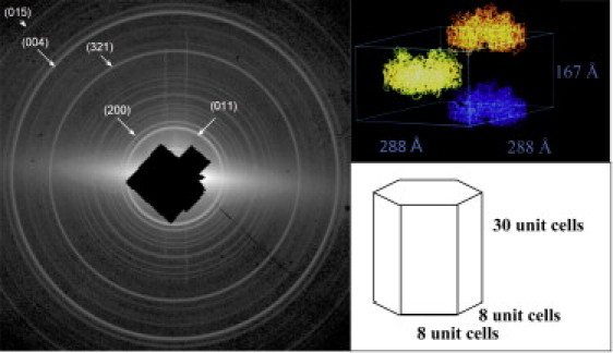
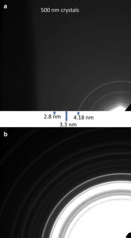
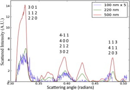
Membrane proteins constitute > 30% of the proteins in an average cell, and yet the number of currently known structures of unique membrane proteins is < 300. To develop new concepts for membrane protein structure determination, we have explored the serial nanocrystallography method, in which fully hydrated protein nanocrystals are delivered to an x-ray beam within a liquid jet at room temperature. As a model system, we have collected x-ray powder diffraction data from the integral membrane protein Photosystem I, which consists of 36 subunits and 381 cofactors. Data were collected from crystals ranging in size from 100 nm to 2 μm. The results demonstrate that there are membrane protein crystals that contain < 100 unit cells (200 total molecules) and that 3D crystals of membrane proteins, which contain < 200 molecules, may be suitable for structural investigation. Serial nanocrystallography overcomes the problem of x-ray damage, which is currently one of the major limitations for x-ray structure determination of small crystals. By combining serial nanocrystallography with x-ray free-electron laser sources in the future, it may be possible to produce molecular-resolution electron-density maps using membrane protein crystals that contain only a few hundred or thousand unit cells.
Copyright © 2011 Biophysical Society. Published by Elsevier Inc. All rights reserved.
Figures


References
-
- Rasmussen S.G.F., Choi H.J., Kobilka B.K. Crystal structure of the human β2 adrenergic G-protein-coupled receptor. Nature. 2007;450:383–387. - PubMed
-
- Rosenbaum D.M., Cherezov V., Kobilka B.K. GPCR engineering yields high-resolution structural insights into β2-adrenergic receptor function. Science. 2007;318:1266–1273. - PubMed
-
- Ago H., Kanaoka Y., Miyano M. Crystal structure of a human membrane protein involved in cysteinyl leukotriene biosynthesis. Nature. 2007;448:609–612. - PubMed
Publication types
MeSH terms
Substances
Grants and funding
LinkOut - more resources
Full Text Sources
Other Literature Sources
