

A mathematical model of the myogenic response to systolic pressure in the afferent arteriole
- PMID: 21190949
- PMCID: PMC3064127
- DOI: 10.1152/ajprenal.00382.2010
A mathematical model of the myogenic response to systolic pressure in the afferent arteriole
Abstract
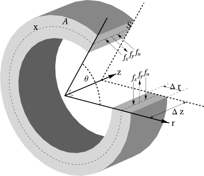
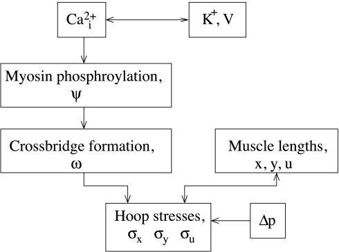
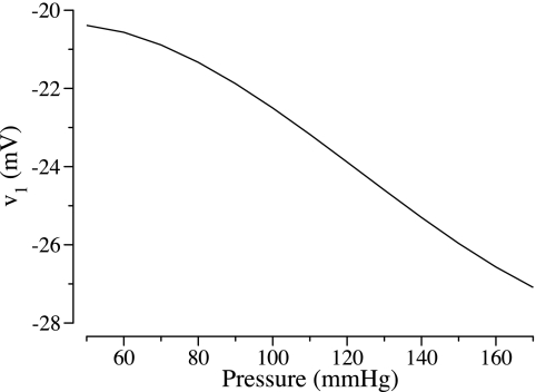
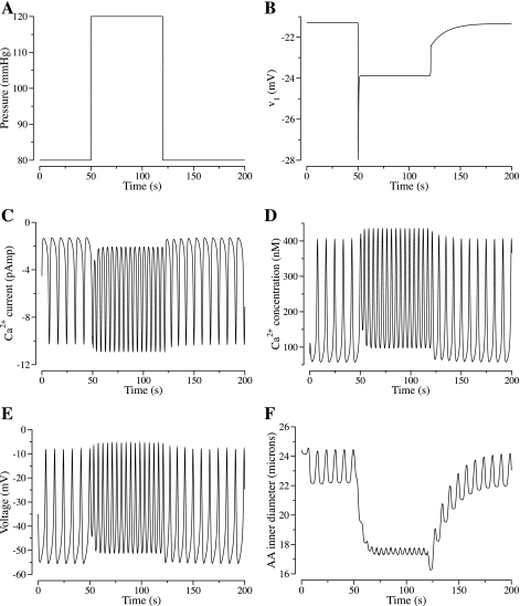
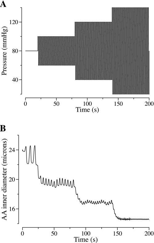
Elevations in systolic blood pressure are believed to be closely linked to the pathogenesis and progression of renal diseases. It has been hypothesized that the afferent arteriole (AA) protects the glomerulus from the damaging effects of hypertension by sensing increases in systolic blood pressure and responding with a compensatory vasoconstriction (Loutzenhiser R, Bidani A, Chilton L. Circ Res 90: 1316-1324, 2002). To investigate this hypothesis, we developed a mathematical model of the myogenic response of an AA wall, based on an arteriole model (Gonzalez-Fernandez JM, Ermentrout B. Math Biosci 119: 127-167, 1994). The model incorporates ionic transport, cell membrane potential, contraction of the AA smooth muscle cell, and the mechanics of a thick-walled cylinder. The model represents a myogenic response based on a pressure-induced shift in the voltage dependence of calcium channel openings: with increasing transmural pressure, model vessel diameter decreases; and with decreasing pressure, vessel diameter increases. Furthermore, the model myogenic mechanism includes a rate-sensitive component that yields constriction and dilation kinetics similar to behaviors observed in vitro. A parameter set is identified based on physical dimensions of an AA in a rat kidney. Model results suggest that the interaction of Ca(2+) and K(+) fluxes mediated by voltage-gated and voltage-calcium-gated channels, respectively, gives rise to periodicity in the transport of the two ions. This results in a time-periodic cytoplasmic calcium concentration, myosin light chain phosphorylation, and cross-bridge formation with the attending muscle stress. Furthermore, the model predicts myogenic responses that agree with experimental observations, most notably those which demonstrate that the renal AA constricts in response to increases in both steady and systolic blood pressures. The myogenic model captures these essential functions of the renal AA, and it may prove useful as a fundamental component in a multiscale model of the renal microvasculature suitable for investigations of the pathogenesis of hypertensive renal diseases.
Figures


References
-
- Bidani AK, Griffin KA. Long-term renal consequenecs of hypertension for normal and diseased kidneys. Curr Opin Nephrol Hypertens 11: 73–80, 2002 - PubMed
-
- Bidani AK, Griffin KA. Pathophysiology of hypertensive renal damage: implications for therapy. Hypertension 44: 1–7, 2004 - PubMed
-
- Cupples WA, Novak P, Novak V, Salevsky FC. Spontaneous blood pressure fluctuations and renal blood flow dynamics. Am J Physiol Renal Fluid Electrolyte Physiol 270: F82–F89, 1996 - PubMed
-
- Davis MJ, Sikes PJ. Myogenic responses of isolated arterioles: test for a rate-sensitive mechanism. Am J Physiol Heart Circ Physiol 259: H1890–H1900, 1990 - PubMed
Publication types
MeSH terms
Substances
Grants and funding
LinkOut - more resources
Full Text Sources
Miscellaneous