. 2011 Mar;85(5):2060-78.
doi: 10.1128/JVI.02210-10. Epub 2010 Dec 29.

Retrovirus-induced spongiform neurodegeneration is mediated by unique central nervous system viral targeting and expression of env alone

Affiliations

Retrovirus-induced spongiform neurodegeneration is mediated by unique central nervous system viral targeting and expression of env alone

Ying Li et al. J Virol. 2011 Mar.

Abstract

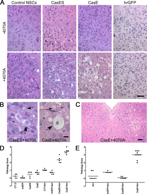
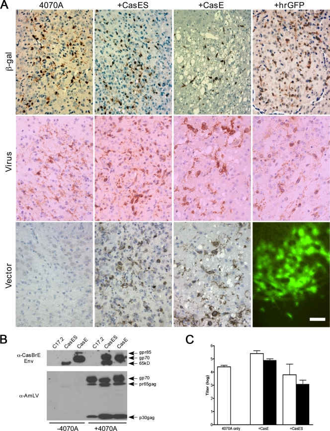
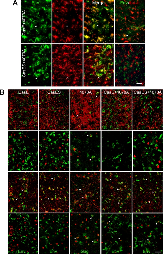
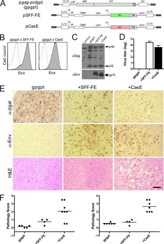
Certain murine leukemia viruses (MLVs) can induce progressive noninflammatory spongiform neurodegeneration similar to that caused by prions. The primary MLV determinants responsible have been mapped to within the env gene; however, it has remained unclear how env mediates disease, whether non-Env viral components are required, and what central nervous system (CNS) cells constitute the critical CNS targets. To address these questions, we examined the effect of transplanting engraftable C17.2 neural stem cells engineered to pseudotype, disseminate, and trans-complement neurovirulent (CasBrE, CasE, and CasES) or non-neurovirulent (Friend and SFF-FE) env sequences (SU or SU/TM) within the CNS using either the "non-neurovirulent" amphotropic helper virus, 4070A, or pgag-polgpt (a nonpackaged vector encoding Gag-Pol). These studies revealed that acute MLV-induced spongiosis results from two separable activities of Env. First, Env causes neuropathology through unique viral targeting within the CNS, which was efficiently mediated by ecotropic Envs (CasBrE and Friend), but not 4070A amphotropic Env. Second, Env induces spongiosis through a toxin activity that is MLV-receptor independent and does not require the coexpression of other viral structural proteins. CasBrE and 4070A Envs possess the toxin activity, whereas Friend Env does not. Although the identity of the critical viral target cell(s) remains unresolved, our results appear to exclude microglia and oligodendrocyte lineage cells, while implicating viral entry into susceptible neurons. Thus, MLV-induced disease parallels prionopathies in that a single protein, Env, mediates both the CNS targeting and the toxicity of the infectious agent that manifests itself as progressive vacuolar neurodegeneration.


Figures

FIG. 1.
FIG. 1.
4070A NSCs pseudotyping CasBrE SU or SU/TM induce acute spongiform neurodegeneration. (A) Representative H&E-stained sections are shown indicative of the histological consequences associated with brain stem transplantation of control, CasES, CasE, and hrGFP NSCs with or without 4070A virus infection (+4070A and −4070A, respectively), at 4 weeks posttransplantation. Bar, 40 μm. (B) High-magnification examples of the spongiform pathology seen in mice receiving CasES+4070A and CasE+4070A NSCs illustrate characteristic vacuolation within the neuropil (small arrows) and the presence of cells possessing intact nuclei but translucent cytoplasm (large arrows). Bar, 10 μm. (C) Example of a focal asymmetric lesion indicative of virus/vector-transducing NSC-mediated changes rather than from recombinant virus entering the CNS through the circulation (see reference 13). Bar, 100 μm. (D and E) A summary of the pathology scores observed for mice receiving NSC transplants within the brain stem (D) or lateral cerebral ventricles (E) is shown for 4 weeks postinjection. 4070A virus-infected NSCs are denoted by “+Am”.
FIG. 2.
FIG. 2.
4070A virus-pseudotyping NSCs engraft and disseminate CasBrE Env vectors within the CNS. (A) Representative paraffin brain sections from mice transplanted with control, CasES, CasE, and hrGFP NSCs plus 4070A after immunostaining for β-galactosidase (β-gal; brown), a genetic marker engineered into C17.2 NSCs (53); total virus expression (Virus; brown); or CasBrE Env expression (Vector; brown/black). hrGFP vector expression was assessed in frozen brain sections by direct examination under epifluorescence illumination (lower right panel). Bar, 40 μm. (B) Semiquantitative immunoblot assessment of the 4070A virus/CasBrE Env protein expression levels from equivalent brain extracts of freshly frozen brain sections from mice transplanted with control, CasES, and CasE NSCs with or without 4070A. Samples were separated on 8% SDS-PAGE gels and immunoblotted with MAb 697 (top) and pig anti-AmLV antiserum (bottom). The results are representative of at least three separate mouse brains for each group and at least three separate samples per brain. (C) Virus titration analysis on 4070A-infected control, CasES, and CasE NSC-transplanted brains for total virus (white bars) and CasBrE Env-encoding virus (black bars). Virus titers were determined on samples taken from at least three separate transplanted brains in each group. Error bars indicate the standard deviations. All analyses were performed on mice sacrificed at 4 weeks after NSC transplantation.
FIG. 3.
FIG. 3.
Pseudotyping NSCs facilitate CasBrE Env vector expression in host microglia and OPCs, but not mature oligodendrocytes. (A) Representative brain stem sections from mice transplanted with CasE+4070A and CasES+4070A NSCs immunostained for CasBrE Env (Env; green) and the microglial marker Iba-1 (Iba-1; red) and then merged to show colocalization. Env-positive microglia were readily detected in areas with or without spongiform pathology, as indicated by arrowheads; however, there were also areas where Env expression was observed with limited or no colocalization to microglia (far right images). Images were obtained by using epifluorescence optics. Bar, 40 μm. (B) Confocal double-immunofluorescence staining of CasE, CasES, 4070A, CasE+4070A, and CasES+4070A NSC-transplanted brains with antibodies specific for the mature oligodendrocyte markers CNPase (red; top row) and carbonic anhydrase II (red; second row); the oligodendrocyte progenitor (OPC) markers NG2 (red; third row) and Olig2 (red; bottom row); and either CasBrE Env (Env; green) in CasE, CasES, CasE+4070A, and CasES+4070A NSC transplants or Gag (Gag; green) in 4070A NSC transplants. Arrowheads (white) indicate examples of cells showing Env or Gag colocalization with an oligodendrocyte cell type-specific marker. Bar, 40 μm.
FIG. 4.
FIG. 4.
4070A NSC pseudotyping of a vector encoding the non-neurovirulent ecotropic Friend Env induces acute spongiform neurodegeneration. (A) Structure of the pSFF retroviral vector encoding the Env from Friend virus clone 57, SFF-FE (51). (B) Representative H&E-stained section from a mouse transplanted with NSCs expressing both the 4070A virus and SFF-FE in the brain stem, at 3 weeks posttransplantation, indicative of the severe spongiosis induced with this virus vector combination. (C) Example of a mouse brain stem transplanted with expressing Friend Env (SFF-FE) alone that was not neuropathogenic. (D) Summary of the neuropathology scores for all of the SFF-FE and SFF-FE+4070A NSC mice receiving brain stem transplants (BS) or cerebral ventricle transplants (V). NSC infection with the 4070A virus is indicated by “Am” in the graph labels. Bar, 40 μm.
FIG. 5.
FIG. 5.
CasBrE Env viral particle pseudotyping reveals hidden 4070A neurovirulence. (A) Structure of the p15-1EIH vector used to facilitate CasBrE SU/TM expression without CasBrE env gene packaging into virions. (B) Cell surface expression of CasBrE Env by FACS analysis on C17.2 NSCs after p15-1EIH transfection and selection that persists upon infection with the 4070A virus (right). (C) Immunoblotting for CasBrE Env (top) and Gag (bottom) of postnuclear cell extracts from C17.2 NSCs with (+) or without (−) 15-1EIH and 4070A shows enhanced Gag expression with CasBrE Env. (D) To demonstrate that the CasBrE Env protein was effectively incorporated into virions, culture supernatants from C17.2 NSCs with (+) or without (−)15-1EIH and 4070A were subjected to ultracentrifugation, and the sedimented fraction was evaluated by immunoblotting for CasBrE Env (top) and Gag (bottom). (E) Capacity of 15-1EIH, 4070A, and 15-1EIH+4070A NSCs to produce virus, as assessed by VTA, using a broadly reactive anti-MLV Env antibody. No CasBrE Env-positive foci were observed when VTAs were specifically assessed. (F) Representative example of a 15-1EIH+4070A NSC-transplanted brain stem showing severe spongiform neuropathology at 3 weeks postinjection. Bar, 40 μm. (G) Example of a 15-1EIH+4070A NSC-transplanted brain stem double-immunostained for 4070A viral Gag (green) and the microglial marker Iba-1 (red). Cells exhibiting colocalization (yellow/orange) are identified with arrowheads. Bar, 40 μm. (H) Neuropathology scores for animals receiving control, 15-1EIH, 4070A, and 15-1EIH+4070A NSCs at 3 weeks after brain stem transplantation. (I) Neuropathology scores at 4 weeks for animals receiving control, 15-1EIH, 4070A, and 15-1EIH+4070A NSCs introduced into lateral brain ventricles. (J) Immunoblot assessment of viral protein expression levels in equivalent brain stem lysates from mice receiving 15-1EIH, 4070A, or 15-1EIH+4070A NSCs using anti-AMLV antiserum. Note the 4070A Env, precursor (pr65), and processed (p30) Gag proteins were expressed in brains transplanted with 4070A virus-expressing NSCs with or without 15-1EIH. The data are representative of samples taken from at least three separate brains in each group. No CasBrE Env protein was detected in samples processed in parallel and immunoblotted with CasBrE Env-specific antibody (MAb 697 [data not shown]).
FIG. 6.
FIG. 6.
Infectious CasBrE env alone is sufficient for inducing spongiform neurodegeneration. (A) Schematic representation of the molecular constructs used to produce packaging C17.2 NSCs that encode either neurovirulent (CasE) or non-neurovirulent (SFF-FE) MLV env's. The pgag-polgpt vector (gpgpt) encodes Gag and Gag-Pol polyproteins but because the psi sequence has been deleted (ΔΨ) the RNA is not incorporated into virions. The pCasE and pSFF-FE vectors possess the psi sequence allowing for packaging of the transcribed RNA into particles. (B) FACS analysis of NSCs transfected with pgag-polgpt (open traces), followed by transduction with viruses encoding SFF-FE (left panel) or CasE (right panel) vectors (gray traces). (C) Gag (αGag) and Env (αEnv) immunoblots of equivalent extracts from the cells shown in panel B. The results are representative of three different analyses. (D) VTAs for CasBrE and Friend Env vectors from control, SFF-FE, and CasE-gpgpt NSCs, respectively. (E) Representative sections from mice receiving P0 brain stem transplants of control (gpgpt, left), SFF-FE (+SFF-FE), and CasE+gpgpt (+CasE) NSCs, taken 3 weeks postinjection, immunostained for β-galactosidase (α-β-gal; brown), Env (α-Env; brown), or stained with H&E. Bar, 100 μm. (F) Summary of the neuropathology scores for mice transplanted with the gpgpt-based packaging NSCs after 3 weeks (left) and 4 weeks (right).
FIG. 7.
FIG. 7.
CasE-gpgpt packaging NSC transplants facilitate minimal CasBrE Env expression in host cells. (A) Double immunofluorescence staining for Env (green) and Iba-1 (red) to assess env vector dissemination to host microglia in brains transplanted with gpgpt+SFF-FE (top) and gpgpt+CasE NSCs (bottom), 3 weeks posttransplantation. Arrowheads indicate cells showing colocalization of the two markers. An arrow indicates an example of an Env+, Iba-1 cell in a gpgpt+SFF-FE brain with a morphology consistent with a transplanted NSC. CasBrE Env+ cells possess a similar morphology. CasBrE Env/Iba-1 double-positive cells were only occasionally noted in the CasE+gpgpt NSC-transplanted mice (inset); however, they were uncommon in areas exhibiting vacuolation, observed in the sections as areas devoid of background tissue fluorescence, denoted by the white circles in gpgpt+CasE. The sections were assessed by using epifluorescence optics. Bar, 40 μm. (B) Confocal images of NSC-transplanted brain sections double immunostained for viral protein (green; Gag or Env) and the cell type-specific markers NG2 and Olig2 (red). White arrowheads indicate examples of viral protein expression in NG2- and Oligo2-positive cells (confirmed by z-stack analysis [data not shown]) that minimally represent the engrafted packaging NSCs. Bar, 20 μm. (C) Confocal images of brain stems transplanted with gpgpt packaging NSCs, double immunostained for Env (top) or Gag (bottom) and the pan-neuronal marker HuC/D (red). Note that no Env expression was associated with HuC/D+ neurons; however, examples of Gag immunostaining coincident with HuC/D were detectable in brain stems transplanted with either FE or CasE+gpgpt NSCs, illustrating particulate staining for Gag that was often clustered within the neuron cell bodies (yellow). Bar, 20 μm.

Similar articles

Cited by

References

    1. Andrews, J. M., and M. B. Gardner. 1974. Lower motor neuron degeneration associated with type C RNA virus infection in mice: neuropathological features. J. Neuropathol. Exp. Neurol. 33:285-307. - PubMed
    1. Askovic, S., F. J. McAtee, C. Favara, and J. L. Portis. 2000. Brain infection by neuroinvasive but avirulent murine oncornaviruses. J. Virol. 74:465-473. - PMC - PubMed
    1. Baszler, T. V., and J. F. Zachary. 1991. Murine retroviral neurovirulence correlates with an enhanced ability of virus to infect selectively, replicate in, and activate resident microglial cells. Am. J. Pathol. 138:655-671. (Erratum, 138:1058.) - PMC - PubMed
    1. Baszler, T. V., and J. F. Zachary. 1990. Murine retroviral-induced spongiform neuronal degeneration parallels resident microglial cell infection: ultrastructural findings. Lab. Invest. 63:612-623. - PubMed
    1. Battini, J. L., J. E. Rasko, and A. D. Miller. 1999. A human cell-surface receptor for xenotropic and polytropic murine leukemia viruses: possible role in G protein-coupled signal transduction. Proc. Natl. Acad. Sci. U. S. A. 96:1385-1390. - PMC - PubMed

Publication types

MeSH terms

Substances