. 2011 Mar;85(6):2723-32.
doi: 10.1128/JVI.02050-10. Epub 2010 Dec 29.

West Nile virus differentially modulates the unfolded protein response to facilitate replication and immune evasion

Affiliations

West Nile virus differentially modulates the unfolded protein response to facilitate replication and immune evasion

Rebecca L Ambrose et al. J Virol. 2011 Mar.

Abstract

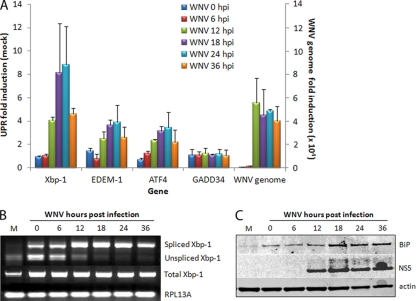
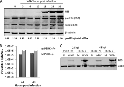
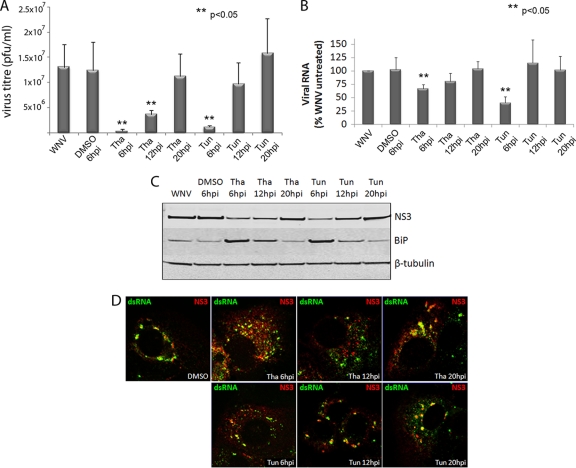
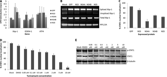
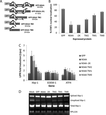
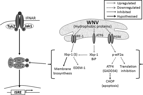
For intracellular survival it is imperative that viruses have the capacity to manipulate various cellular responses, including metabolic and biosynthetic pathways. The unfolded protein response (UPR) is induced by various external and internal stimuli, including the accumulation of misfolded proteins in the endoplasmic reticulum (ER). Our previous studies have indicated that the replication and assembly of the flavivirus West Nile virus strain Kunjin virus (WNV(KUN)) is intimately associated with the ER. Thus, we sought to determine whether the UPR was induced during WNV(KUN) infection. WNV(KUN) induces UPR signaling during replication, which is coordinated with peak replication. Interestingly, signaling is biased toward the ATF6/IRE-1 arm of the response, with high levels of Xbp-1 activation but negligible eukaryotic translation initiation factor 2α phosphorylation and downstream transcription. We show that the PERK-mediated response may partially regulate replication, since external UPR stimulation had a limiting effect on early replication events and cells deficient for PERK demonstrated increased replication and virus release. Significantly, we show that the WNV(KUN) hydrophobic nonstructural proteins NS4A and NS4B are potent inducers of the UPR, which displayed a high correlation in inhibiting Jak-STAT signaling in response to alpha interferon (IFN-α). Sequential removal of the transmembrane domains of NS4A showed that reducing hydrophobicity decreased UPR signaling and restored IFN-α-mediated activation. Overall, these results suggest that WNV(KUN) can stimulate the UPR to facilitate replication and that the induction of a general ER stress response, regulated by hydrophobic WNV(KUN) proteins, can potentiate the inhibition of the antiviral signaling pathway.


Figures

FIG. 1.
FIG. 1.
WNVKUN induces UPR signaling. Vero C1008 cells were infected with WNVKUN at an MOI of 3, and samples collected at 0, 6, 12, 18, 24, and 36 hpi. (A) RNA extracted from infected cells was quantified for upregulation of UPR genes Xbp-1, EDEM-1, ATF4, and GADD34 by using qPCR, and the fold induction was calculated compared to mock cells at the same time point. Error bars indicate +1 standard deviations from replicate assays of two independent experiments. (B) RNA samples from above were also analyzed for spliced Xbp-1 mRNA by using RT-PCR and endonuclease digestion as previously described (58). (C) Protein samples from the time course were analyzed by Western blotting for the synthesis of BiP compared to the infection control NS5 and the internal control actin.
FIG. 2.
FIG. 2.
WNVKUN does not induce or require PERK-mediated signaling for replication. (A) Vero C1008 cells were infected with WNVKUN at an MOI of 3, and protein samples collected at 0, 6, 12, 18, 24, and 36 hpi. Cell lysates were analyzed by Western blotting for p-eIF2α (Ser 51), total eIF2α, NS5, and the internal control tubulin. The ratio of p-eIF2α to eIF2α was quantified by using Bio-Rad Quantity One software. (B) PERK−/− and PERK+/+ MEFs were infected with WNVKUN at an MOI of 3 for 24 and 48 h, and supernatant and protein samples were collected. (C) Plaque assays were used to analyze the release of infectious virus, and Western blotting for NS5 was used to detect viral protein synthesis. Error bars indicate +1 standard deviations for replicate assays of two independent experiments.
FIG. 3.
FIG. 3.
Synthetic UPR activation during WNVKUN infection inhibits replication. Vero C1008 cells were infected with WNVKUN at an MOI of 3 and then treated with 300 nM tunicamycin, thapsigargin, or the drug vehicle (DMSO) at 6, 12, or 20 hpi. At 24 hpi, viral supernatants, protein, and RNA samples were collected, as well as fixed for immunofluorescence, and analyzed for viral replication. (A and B) Virus titers were determined by plaque assays (A), and RNA samples were analyzed for viral RNA expression by qPCR (compared to internal control RPL13A) (B). (C) Cell protein lysates were analyzed for viral protein expression and BiP upregulation by Western blotting, compared to the internal control actin. (D) Cells were fixed using acetone-methanol and labeled for NS3 (in red) and dsRNA (in green) to study formation of replication complexes after treatment. Error bars indicate +1 standard deviations of two independent experiments. Statistical analysis was performed by using the Student t test.
FIG. 4.
FIG. 4.
WNV NS proteins differentially regulate UPR and IFN signaling. Vero C1008 cells were transfected with GFP fusion constructs of NS3, NS4A, NS4B, and NS5 and expressed for 18 h. The cells were then sorted according to GFP expression, and a pool of GFP-positive cells was collected. A total of 5 × 10⁴ cells were lysed, and extracted RNA was analyzed for the upregulation of UPR genes Xbp-1, EDEM-1, and ATF4 (A) and the presence of spliced Xbp-1 mRNA (B). Error bars indicate +1 standard deviations of replicate assays of two independent experiments. (C) Cells were also transfected with the above constructs, stimulated with 1,000 U of IFN-α (Roche)/ml at 24 h posttransfection (hpt) for 30 min, and then fixed and labeled with STAT1 and DAPI (4′,6′-diamidino-2-phenylindole) to detect nuclear trafficking. Approximately 100 expressing cells per sample were scored on nuclear or cytoplasmic localization. Error bars indicate +1 standard deviations from two independent experiments. Vero C1008 cells were treated with different concentrations of tunicamycin for 12 h at 37°C and then stimulated with 1,000 U of IFN-α (Roferon; Roche)/ml for 30 min at the specified tunicamycin concentration. The cells were then fixed for immunofluorescence or lysed for Western blot analysis. (D) Fixed cells were labeled with a STAT1 antibody, and the nuclear stain DAPI to detect nuclear localization. Approximately 100 cells per concentration were quantified for nuclear localization. Error bars indicate +1 standard deviations from two independent experiments. (E) Cell lysates were probed for phospho-STAT1 and STAT1 to detect STAT1 phosphorylation and compared to the internal control actin.
FIG. 5.
FIG. 5.
Deletion of TM regions from NS4A results in decreased regulation of UPR and IFN signaling. (A) TM deletion mutants of NS4A were created (based upon TM predictions by Miller et al. [38]) with GFP at the N terminus. Vero C1008 cells were transfected with full-length NS4A and the TM mutants (as well as a GFP control). (B) At 24 hpt, the cells were fixed and labeled with STAT1 and the nuclear stain DAPI to detect nuclear localization in expressing cells. Approximately 100 cells for each fusion protein were scored for STAT localization as either complete nuclear, partial nuclear, or cytoplasmic localization, and percentage nuclear trafficking calculated. Error bars indicate +1 standard deviations from duplicate experiments. (C) At 18 hpt, cells were collected in MACS buffer and sorted by using FACS to create a pool of expressing cells, and then 5 × 10⁴ cells were lysed for RNA extraction and qPCR analyses. RNA was analyzed for the upregulation of Xbp-1, EDEM-1, and ATF4 compared to the internal control RPL13A. Error bars indicate +1 standard deviations from duplicate experiments. (D) RNA samples were also analyzed for splicing of the Xbp-1 mRNA.
FIG. 6.
FIG. 6.
WNV replication and/or hydrophobic NS protein expression regulates the UPR in order to inhibit Jak-STAT signaling. WNV replication and/or expression of hydrophobic proteins (NS2B, NS4A, and NS4B) regulates UPR signaling to benefit replication. The ATF6/IRE-1 pathways are strongly activated, resulting in Xbp-1 transcription and splicing which may aid in the proliferation of ER membranes for the replication complex. In contrast, the PERK arm of the UPR is not strongly induced, potentially avoiding inhibitory downstream effects, such as translation inhibition and apoptosis, which may limit replication. Downstream UPR signaling during WNV replication can also inhibit STAT1 phosphorylation and nuclear trafficking; however, this is dependent on expression of hydrophobic proteins, since the removal of hydrophobic domains resulted in decreased Xbp-1 activation and the restoration of STAT1 signaling.

References

    1. Anthony, T. G., B. C. McGrath, D. R. Cavener, and R. C. Wek. 2003. The eIF2a kinase GCN2 is necessary for adaptation to dietary essential amino acid (EAA) deprivation in growing mice. FASEB J. 17:A811-A811.
    1. Ashour, J., M. Laurent-Rolle, P. Y. Shi, and A. Garcia-Sastre. 2009. NS5 of dengue virus mediates STAT2 binding and degradation. J. Virol. 83:5408-5418. - PMC - PubMed
    1. Baltzis, D., et al. 2004. Resistance to vesicular stomatitis virus infection requires a functional cross talk between the eukaryotic translation initiation factor 2 alpha kinases PERK and PKR. J. Virol. 78:12747-12761. - PMC - PubMed
    1. Bernales, S., F. R. Papa, and P. Walter. 2006. Intracellular signaling by the unfolded protein response. Annu. Rev. Cell Dev. Biol. 22:487-508. - PubMed
    1. Best, S. M., et al. 2005. Inhibition of interferon-stimulated JAK-STAT signaling by a tick-borne flavivirus and identification of NS5 as an interferon antagonist. J. Virol. 79:12828-12839. - PMC - PubMed

Publication types

MeSH terms

LinkOut - more resources