. 2011 Mar;85(6):2611-9.
doi: 10.1128/JVI.02080-10. Epub 2010 Dec 29.

Differential effects of human immunodeficiency virus type 1 Vpu on the stability of BST-2/tetherin

Affiliations

Differential effects of human immunodeficiency virus type 1 Vpu on the stability of BST-2/tetherin

Amy J Andrew et al. J Virol. 2011 Mar.

Abstract

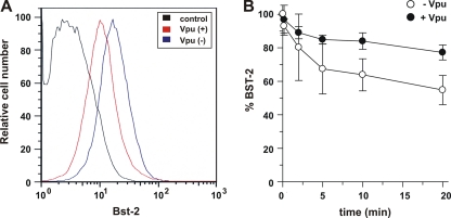
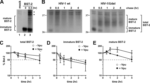
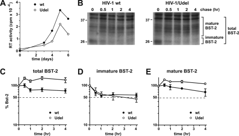
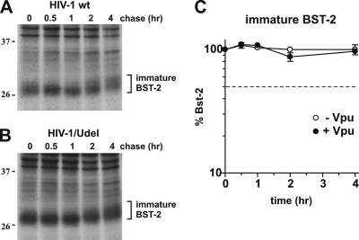
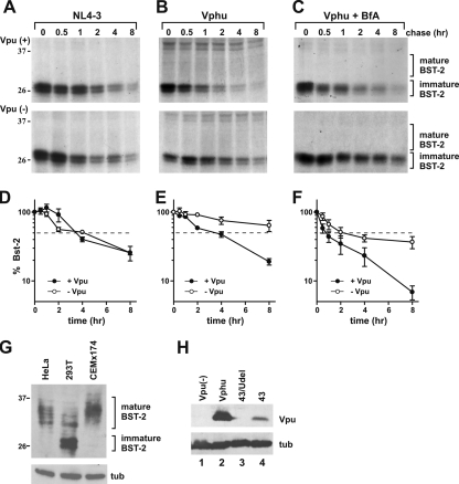
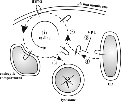
BST-2/CD317/tetherin is a host factor that inhibits the release of HIV-1 and other unrelated viruses. A current model proposes that BST-2 physically tethers virions to the surface of virus-producing cells. The HIV-1-encoded Vpu protein effectively antagonizes the activity of BST-2. How Vpu accomplishes this task remains unclear; however, it is known that Vpu has the ability to down-modulate BST-2 from the cell surface. Here we analyzed the effects of Vpu on BST-2 by performing a series of kinetic studies with HeLa, 293T, and CEMx174 cells. Our results indicate that the surface downregulation of BST-2 is not due to an accelerated internalization or reduced recycling of internalized BST-2 but instead is caused by interference with the resupply of newly synthesized BST-2 from within the cell. While our data confirm previous reports that the high-level expression of Vpu can cause the endoplasmic reticulum (ER)-associated degradation of BST-2, we found no evidence that Vpu targets endogenous BST-2 in the ER in the course of a viral infection. Instead, we found that Vpu acts in a post-ER compartment and increases the turnover of newly synthesized mature BST-2. Our observation that Vpu does not affect the recycling of BST-2 suggests that Vpu does not act directly at the cell surface but may interfere with the trafficking of newly synthesized BST-2 to the cell surface, resulting in the accelerated targeting of BST-2 to the lysosomal compartment for degradation.


Figures

FIG. 1.
FIG. 1.
Vpu reduces BST-2 levels independently of the rate of endocytosis of BST-2. (A) HeLa cells were transfected with 1 μg of pEGFP-N1 with [Vpu(+)] or without [Vpu(−)] 1 μg of pcDNA-Vphu. The total DNA was adjusted to 5 μg with empty vector DNA. Cells were stained with BST-2 antibodies and analyzed with a FACSCalibur instrument. Samples were gated for GFP expression, which was used as a marker for transfected cells. As a control, HeLa cells were stained with preimmune serum (control). (B) Cells were transfected as described above for A, harvested 24 h after transfection, labeled at 4°C with antibody to BST-2, and then incubated at 37°C for the indicated times. Cells were then stained with a secondary antibody and analyzed with a FACSCalibur instrument. Samples were gated for GFP-positive cells. The mean fluorescence intensity of the time zero cells for each population was defined as 100% BST-2 surface expression. Plotted data were derived from five independent experiments. Error bars represent standard errors of the means.
FIG. 2.
FIG. 2.
Vpu accelerates the turnover of mature endogenous BST-2 in HeLa cells. (A) 293T cells were transfected with 1 μg of Bst-2, pulse-labeled for 30 min by the addition of 35S-labeled amino acids, and chased for 30 min in complete medium. Mock represents untransfected cells labeled and immunoprecipitated in a similar manner. (B) HeLa cells were infected with pCMV-VSVg-pseudotyped pNL4-3 or pNL4-3/Udel virus along with 8 μg/μl polybrene. Twenty-four hours after infection cells were pulse-labeled for 1 h at 37°C by addition of 35S-labeled amino acids and chased for the indicated time intervals in complete medium. BST-2 was immunoprecipitated with specific antibodies. A representative result is shown. The position of molecular weight standards is shown on the left. (C) The percentage of BST-2 remaining at each time point was calculated relative to the signal at time zero. (D and E) Total levels of BST-2 were calculated by adding both immature BST-2 (D) and mature BST-2 (E). Mature BST-2 and immature BST-2 are indicated on the right. Plotted data are derived from three independent experiments. Error bars represent standard errors of the means.
FIG. 3.
FIG. 3.
Vpu accelerates the turnover of mature endogenous BST-2 in CEMx174 cells. (A) CEMx174 cells were infected with equal reverse transcriptase (RT) units of wt NL4-3 and NL4-3/Udel virus stocks produced in 293T cells. Virus replication was monitored by measuring the virus-associated reverse transcriptase activity in culture supernatants as described previously (44). (B) On day 5 after infection cells were pulse-labeled for 1 h at 37°C by the addition of 35S-labeled amino acids and chased for the indicated time intervals in complete medium. BST-2 was immunoprecipitated with specific antibodies. A representative experiment is shown. Mature BST-2 and high-mannose BST-2 are indicated. The position of molecular weight standards is shown on the left. (C) The percentage of BST-2 remaining at each time point was calculated relative to the signal at time zero. (D and E) Total levels of BST-2 were calculated by adding both immature BST-2 (D) and mature BST-2 (E). Plotted data are derived from three independent experiments. Error bars represent standard errors of the means.
FIG. 4.
FIG. 4.
(A and B) CEMx174 cells were infected with equal reverse transcriptase units of wt NL4-3 (A) and NL4-3/Udel (B) virus stocks produced in 293T cells as described in the legend of Fig. 3. On day 5 after infection cells were pulse-labeled as described in the legend of Fig. 3, with the addition of BfA during the pulse and chase at 5 μg/ml. A representative result is shown. The position of molecular weight standards is shown on the left. (C) Graph comparing the levels of the immature form of BST-2 from cells infected by wt HIV-1 (closed circles) and HIV-1/Udel (open circles). Plotted data are derived from three independent experiments. Error bars represent standard errors of the means.
FIG. 5.
FIG. 5.
Transfected BST-2 in 293T cells is destabilized by VphU but not Vpu. (A) 293T cells were transfected with 1 μg of pCDNA-BST-2 with 4 μg of either wt NL4-3 virus (closed circles) or NL4-3/Udel virus (open circles). (B) 293T cells were transfected with 1 μg of pcDNA-BST2 with (closed circles) or without (open circles) 2 μg pcDNA-VphU. Total DNA was adjusted to 5 μg with empty vector DNA. (C) 293T cells were transfected as described above (B), with the addition of brefeldin A during the pulse and chase at 5 μg/ml. In all experiments, 24 h after transfection cells were pulse-labeled for 1 h at 37°C by the addition of 35S-labeled amino acids and chased for the indicated time intervals in complete medium. BST-2 was immunoprecipitated with specific antibodies. Representative results are shown. The position of molecular weight standards is shown on the left. (D to F) The percentage of immature BST-2 remaining at each time point was calculated relative to the signal at time zero. Plotted data are derived from three independent experiments. Error bars represent standard errors of the means. (G) Cell lysates from 293T cells transfected with 1 μg of BST-2 as well as HeLa and CEMx174 cell extracts were separated by SDS-PAGE and subjected to immunoblot analysis with antibodies to BST-2 and tubulin (tub). The position of molecular weight standards is shown on the left. (H) 293T cells were transfected with 1 μg of pcDNA-BST2 in the absence of Vpu (lane 1) or together with 2 μg of pcDNA-VphU (lane 2), 4 μg of pNL4-3/Udel (lane 3), or 4 μg of pNL4-3 DNA (lane 4). Total DNA was adjusted to 5 μg with empty vector DNA. Lysates were separated by SDS-PAGE and subjected to immunoblot analysis with antibodies to Vpu, or tubulin (tub).
FIG. 6.
FIG. 6.
Model for the interference of Vpu with cell surface expression of BST-2. BST-2 expressed at the cell surface is not static but cycles between the endocytic compartment and the cell surface (1). The majority of internalized BST-2 (as indicated by the size of the arrowhead) is recycled to the cell surface (2). A small fraction of internalized BST-2 molecules is not recycled to the surface but remains inside the cell and is targeted for degradation in the lysosomal compartment (3). The BST-2 population at the cell surface is maintained at constant levels by replacing the nonrecycled portion of BST-2 with de novo-synthesized protein from the endoplasmic reticulum (ER) (4). The interference of Vpu with the resupply (5) results in the gradual depletion of cell surface BST-2.

References

    1. Adachi, A., et al. 1986. Production of acquired immunodeficiency syndrome-associated retrovirus in human and nonhuman cells transfected with an infectious molecular clone. J. Virol. 59:284-291. - PMC - PubMed
    1. Andrew, A., and K. Strebel. 2010. HIV-1 Vpu targets cell surface markers CD4 and BST-2 through distinct mechanisms. Mol. Aspects Med. 31:407-417. - PMC - PubMed
    1. Andrew, A. J., E. Miyagi, S. Kao, and K. Strebel. 2009. The formation of cysteine-linked dimers of BST-2/tetherin is important for inhibition of HIV-1 virus release but not for sensitivity to Vpu. Retrovirology 6:80. - PMC - PubMed
    1. Asaoka, K., et al. 2005. A retrovirus restriction factor TRIM5alpha is transcriptionally regulated by interferons. Biochem. Biophys. Res. Commun. 338:1950-1956. - PubMed
    1. Bartee, E., A. McCormack, and K. Fruh. 2006. Quantitative membrane proteomics reveals new cellular targets of viral immune modulators. PLoS Pathog. 2:e107. - PMC - PubMed

Publication types

MeSH terms

Substances

LinkOut - more resources