. 2011 Feb 15;519(3):420-32.
doi: 10.1002/cne.22525.

Serotonin receptor diversity in the human colon: Expression of serotonin type 3 receptor subunits 5-HT3C, 5-HT3D, and 5-HT3E

Affiliations

Serotonin receptor diversity in the human colon: Expression of serotonin type 3 receptor subunits 5-HT3C, 5-HT3D, and 5-HT3E

Johannes Kapeller et al. J Comp Neurol. .

Abstract

Since the first description of 5-HT₃ receptors more than 50 years ago, there has been speculation about the molecular basis of their receptor heterogeneity. We have cloned the genes encoding novel 5-HT3 subunits 5-HT3C, 5-HT3D, and 5-HT3E and have shown that these subunits are able to form functional heteromeric receptors when coexpressed with the 5-HT3A subunit. However, whether these subunits are actually expressed in human tissue remained to be confirmed. In the current study, we performed immunocytochemistry to locate the 5-HT3A as well as the 5-HT3C, 5-HT3D, and 5-HT3E subunits within the human colon. Western blot analysis was used to confirm subunit expression, and RT-PCR was employed to detect transcripts encoding 5-HT₃ receptor subunits in microdissected tissue samples. This investigation revealed, for the first time, that 5-HT3C, 5-HT3D, and 5-HT3E subunits are coexpressed with 5-HT3A in cell bodies of myenteric neurons. Furthermore, 5-HT3A and 5-HT3D were found to be expressed in submucosal plexus of the human large intestine. These data provide a strong basis for future studies of the roles that specific 5-HT₃ receptor subtypes play in the function of the enteric and central nervous systems and the contribution that specific 5-HT₃ receptors make to the pathophysiology of gastrointestinal disorders such as irritable bowel syndrome and dyspepsia.


Figures

Figure 1
Figure 1
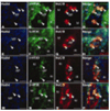
Immunocytochemical analysis of the immunoreactivity of 5-HT3 subunits within the myenteric plexus of the human colon. The immunoreactivities of the 5-HT3C (A–D), 5-HT3D (E–H), and 5-HT3E (I–L ) subunits (green fluorescence, fluorescein) are coincident (arrowheads) with the immunoreactivity of the 5-HT3A subunit (red fluorescence, tetramethylrhodamine) in the perikarya of neurons of the myenteric plexus (MP) within the muscular layer (M). Blue staining marks nuclei (DAPI counterstain). Scale bars = 100 µm.
Figure 2
Figure 2
Immunocytochemical analysis of the immunoreactivity of 5-HT3 subunits within the submucosal plexus of the human colon. The immunoreactivities of the 5-HT3A (A–D) and 5-HT3D (E–H) subunits (green fluorescence, fluorescein) are coincident (arrowheads) with the neuronal marker staining (HuC/D; red fluorescence) in the perikarya of neurons of the submucosal plexus (SMP). Blue staining marks nuclei (DAPI counterstain). Scale bars = 50 µm.
Figure 3
Figure 3
Confirmation of HTR3 genes expression by RT-PCR from microdissected colon tissue. Microdissected tissue of the mucosal cell layer of the epithelium and of the muscular layer of the human colon was analyzed by RT-PCR. All tested HTR3 genes are expressed within enterocytes (E) as well as in the lamina propria (LP). In contrast, in the muscular layer (M), only a weak expression of HTR3D is detectable whereas there is expression of HTR3A, HTR3C, HTR3D, and HTR3E in myenteric plexus (MP). CK20, cytokeratin 20; CD45, protein tyrosine phosphatase, receptor type C; ACTG2, actin, gamma 2; ELAVL13, HuC/D; ARF, ADP-ribosylation factor; NC, negative control.
Figure 4
Figure 4
Colon colocalization studies of 5-HT3A, 5-HT3C, 5-HT3D, and 5-HT3E with HuC/D. 5-HT3A (A–D), 5-HT3C (E–H), 5-HT3D (I–L ), and 5-HT3E (M–P) are expressed in HuC/D-positive cell somata of neurons of the myenteric plexus (MP) in the muscular layer (M) of the colon. Coexpression is indicated by arrowheads. 5-HT3 subunits are labeled in green, and the neuronal marker HuC/D is stained in red. Blue staining marks nuclei (DAPI counterstain). Scale bars = 50 µm.
Figure 5
Figure 5
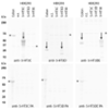
Western blot analysis of a colon tissue lysate. Shown are results obtained with a colon cell lysate of one individual compared with lysates from HEK293 cells transiently transfected with the respective 5-HT3 subunit (positive control) and nontransfected cells (n.t., as negative control). Immunoreactive bands in the colon sample were detectable for 5-HT3D of ~25 kDa and for 5-HT3E of ~40–45 kDa (top). Specificity of antibodies was shown by preadsorption (PA) of the primary antibodies with the respective antigen peptide (bottom). Arrows indicate specific bands; asterisks indicate nonspecific background bands.
Figure 6
Figure 6
Schematic illustration of 5-HT3 subunit expression in the human colon. Shown are the summarized results of this study as well as the previously described 5-HT3B expression within the SMP (Michel et al., 2005). Basic organization of this figure was adapted from Mawe et al. (1986). DRG, dorsal root ganglion; MP, myenteric plexus; SMP, submucosal plexus.

Similar articles

Cited by

References

    1. Barnes NM, Hales TG, Lummis SC, Peters JA. The 5-HT3 receptor—the relationship between structure and function. Neuropharmacology. 2009;56:273–284. - PMC - PubMed
    1. Bearcroft CP, Andre EA, Farthing MJ. In vivo effects of the 5-HT3 antagonist alosetron on basal and cholera toxin-induced secretion in the human jejunum: a segmental perfusion study. Aliment Pharmacol Ther. 1997;11:1109–1114. - PubMed
    1. Belelli D, Balcarek JM, Hope AG, Peters JA, Lambert JJ, Blackburn TP. Cloning and functional expression of a human 5-hydroxytryptamine type 3AS receptor subunit. Mol Pharmacol. 1995;48:1054–1062. - PubMed
    1. Bertrand PP, Kunze WAA, Bornstein JC, Furness JB, Smith ML. Analysis of the responses of myenteric neurons in the small intestine to chemical stimulation of the mucosa. Am J Physiol. 1997;36:G422–G435. - PubMed
    1. Bottner M, Bar F, Von Koschitzky H, Tafazzoli K, Roblick UJ, Bruch HP, Wedel T. Laser microdissection as a new tool to investigate site-specific gene expression in enteric ganglia of the human intestine. Neurogastroenterol Motil. 2009;22:168–172. e152. - PubMed

Publication types