. 2010 Dec 30:10:122.
doi: 10.1186/1471-213X-10-122.

Epithelial cell-directed efferocytosis in the post-partum mammary gland is necessary for tissue homeostasis and future lactation

Affiliations

Epithelial cell-directed efferocytosis in the post-partum mammary gland is necessary for tissue homeostasis and future lactation

Melissa Sandahl et al. BMC Dev Biol. .

Abstract

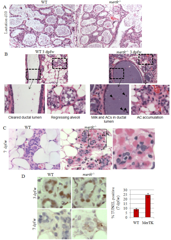
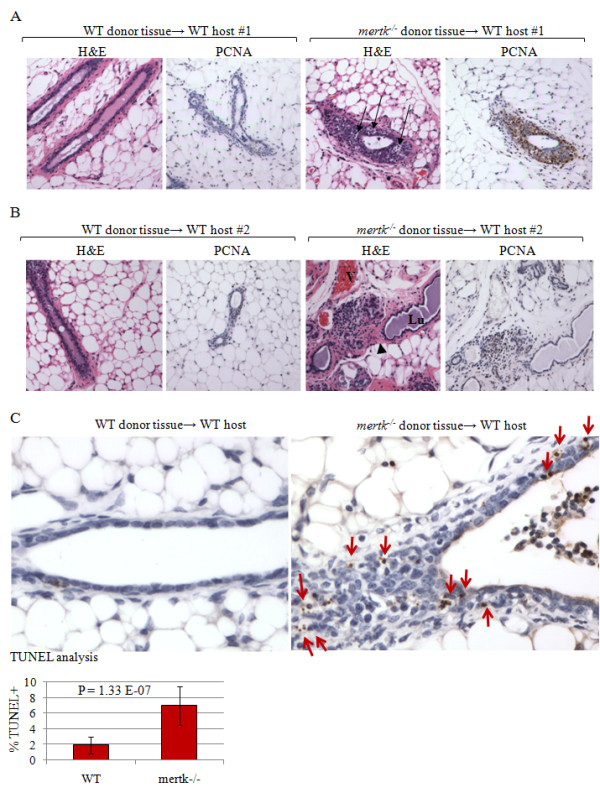
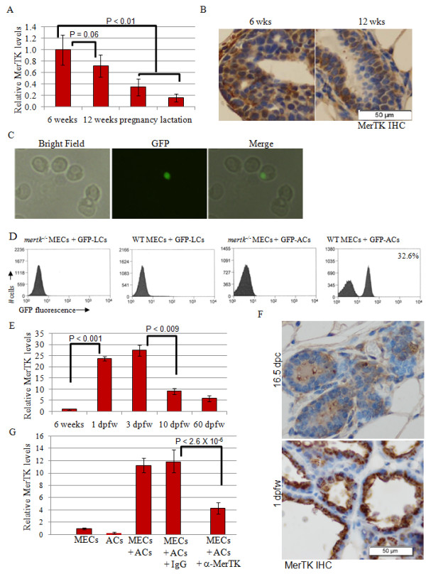
Background: Mammary glands harbor a profound burden of apoptotic cells (ACs) during post-lactational involution, but little is known regarding mechanisms by which ACs are cleared from the mammary gland, or consequences if this process is interrupted. We investigated AC clearance, also termed efferocytosis, during post-lactational remodeling, using mice deficient for MerTK, Axl, and Tyro3, three related receptor tyrosine kinases (RTKs) regulating macrophage-mediated efferocytosis in monocytes. MerTK expression, apoptosis and the accumulation of apoptotic debris were examined in histological sections of MerTK-deficient, Axl/Tyro3-deficient, and wild-type mammary glands harvested at specific time points during lactation and synchronized involution. The ability of primary mammary epithelial cells (MECs) to engulf ACs was assessed in culture. Transplant of MerTK-deficient mammary epithelium into cleared WT mammary fat pads was used to assess the contribution of WT mammary macrophages to post-lactational efferocytosis.

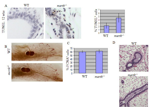
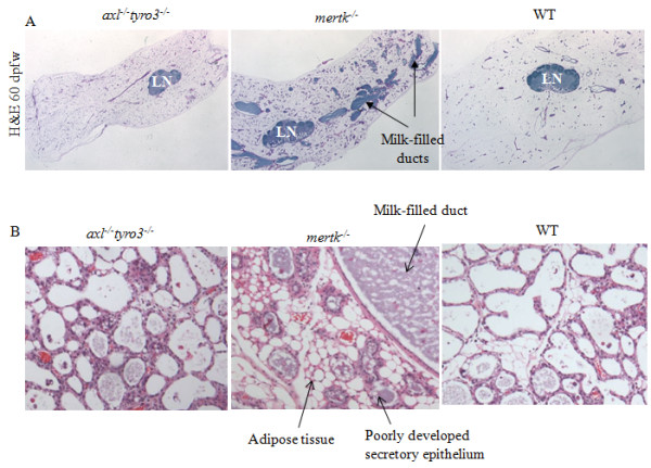
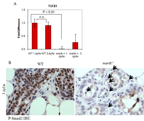
Results: ACs induced MerTK expression in MECs, resulting in elevated MerTK levels at the earliest stages of involution. Loss of MerTK resulted in AC accumulation in post-lactational MerTK-deficient mammary glands, but not in Axl and Tyro3-deficient mammary glands. Increased vascularization, fibrosis, and epithelial hyperproliferation were observed in MerTK-deficient mammary glands through at least 60 days post-weaning, due to failed efferocytosis after lactation, but did not manifest in nulliparous mice. WT host-derived macrophages failed to rescue efferocytosis in transplanted MerTK-deficient mammary epithelium.

Conclusion: Efferocytosis by MECs through MerTK is crucial for mammary gland homeostasis and function during the post-lactational period. Efferocytosis by MECs thus limits pathologic consequences associated with the apoptotic load following lactation.


Figures

Figure 1
Figure 1
Impaired clearance of apoptotic mammary epithelial cells during involution in MerTK-deficient mice. A. H&E-stained sections of mammary glands harvested at lactation day 10 from WT or mertk-/- mammary glands. B-C. H&E-stained sections of mammary glands harvested at 3 dpfw (B) and 7 dpfw (C) from WT and mertk-/- mammary glands. Boxed areas are shown in higher magnification, where indicated in B indicate AC accumulation in the ductal lumen and in regressing alveolar structures. Arrows in C indicate apoptotic cells surrounding live cells in a rosette pattern. D Immunohistochemical detection of TUNEL-positive cells at 7 dpfw. Values shown in D (left panel) are the average percentage of total nuclei that are TUNEL-positive. N = 5 (X 3 random 400X fields per sample). P = 0.002, Student's unpaired T-test.
Figure 2
Figure 2
Impaired apoptotic cell clearance results in pathological changes that are sustained through 60 dpfw. A. Sections of mammary glands harvested at 60 dpfw were stained with H&E and with an antibody against collagen. B. Immunohistochemical detection of PCNA in mammary glands harvested at 60 dpfw. N = 5 per genotype. Values shown in bottom left of each panel represent the average percentage of total nuclei per 400X field (± S.D.) that were PCNA+. *P = 0.045; Student's unpaired T-test, N = 3 samples per group, 5 fields per sample. C. Immunohistochemical detection of the blood vessel marker PECAM/CD31. Values shown in the bottom left of each panel represent the average number of vCD31-positive vessels per 400X field (± S.D.). **P = 0.0001. Student's unpaired T-test, N = 3 samples per group, 5 fields per sample.
Figure 3
Figure 3
Pathologic consequences of impaired apoptotic cell clearance are specific to the post-partum mammary gland. A. Increased presence of apoptotic cells in MerTK-deficient mice was shown by TUNEL analysis of mammary glands from 12-week old virgin female mice. Values represent the average percentage of the total epithelial population that was TUNEL-positive, ± S.D. N = 3 (5 fields per sample). P = 0.01, Student's T-test. B. Whole mounted, hematoxylin-stained mammary glands harvested from eight week old mice. C. mammary glands from 12-wekk old virgin female mice were analyzed by immunohistochemistry for PCNA. Values shown represent the average percentage of total epithelial nuclei that were PCNA-positive, ± S.D. P = 0.39, Student's T-test. D. H&E-stained sections from WT and mertk-/- mammary glands harvested from virgin mice at 12 weeks of age.
Figure 4
Figure 4
Loss of MerTK, but not Axl or Tyro3, causes post-lactational pathologies that prevent lactation following second pregnancy. A. Low-power magnification of H&E-stained section of WT, mertk-/- and Axl-/-Tyro3-/- mammary glands harvested at 60 dpfw, demonstrating the tissue-wide extent of the pathological changes occurring in MerTK-deficient samples. Arrows indicate milk-filled distended lumens in mertk-/- samples. B. At 60 dpfw, mice were impregnated for a second time. Mammary glands shown here were harvested at lactation day 1. Note the incomplete development of the lobuloalveolar epithelium in MerTK-deficient samples.
Figure 5
Figure 5
WT macrophages do not compensate for impaired efferocytosis by MerTK-deficient mammary epithelium. A-B. Histological examination of WT mammary glands cleared of endogenous epithelium and central lymph nodes and reconstituted with either WT or MerTK-deficient mammary epithelium. For each host mouse, WT tissue was transplanted into the right #4 fat pad, and mertk-/- tissue was transplanted into the left #4 fat pad. Paired sets are displayed. Mice were impregnated 8 weeks following mammary transplant, pups were withdrawn at lactation day 1 to enforce involution, and mammary glands were harvested at 14 dpfw. H&E images and PCNA immunohistochemistry are shown. A. Images taken of the right and left reconstituted mammary glands from host female #1. B. Images taken of the right and left reconstituted mammary glands from host female #2. C. TUNEL analysis of paired reconstituted mammary samples from a single WT recipient mouse, showing the mammary gland reconstituted with WT mammary epithelium and mertk-/- mammary epithelium. Arrows indicate apoptotic bodies. Values represent the average percentage of the total epithelial nuclei that were TUNEL-positive, ± S.D. Significance calculated using Student's paired T-test. N = 7.
Figure 6
Figure 6
MerTK directs efferocytosis in mammary epithelial cells. A. Taqman™ real-time PCR was used to quantitate the relative levels of mertk mRNA in mouse mammary total RNA, harvested at the indicated time points. The relative levels of mertk were calculated using expression of mouse GAPDH to correct for total RNA concentration using the δδCT method, setting the value of mertk expression measured at 6 weeks to 1. Average values are shown ± S.D. N = 3, each sample analyzed 6 times. Statistical significance using Student's T-test (unpaired). B. Immunohistochemistry to detect MerTK in WT mammary glands harvested from virgin female mice at 6 and 12 weeks of age. Representative images are shown. C. WT and mertk-/- PMECs were incubated for 4 hours in the presence of live or apoptotic GFP+ cells harvested from the thymus of untreated or dexamethasone-treated (respectively) actin-GFP mice. Representative photomicrographs of WT PMECs ingesting GFP+ apoptotic thymocytes are shown. D. Flow cytometric analysis of WT and mertk-/- PMECs incubated for 4 hours in the presence of apoptotic cells (ACs) or live cells (LCs). N> 30,000 cells examined per genotype. Value in right panel indicates percentage of total cells that were GFP+. E. Real-time RT-PCR was used to measure mertk mRNA expression in whole mammary RNA harvested from 6 weeks virgin female mice, or from female mice at 1, 3, 10, and 60 dpfw. Values shown represent the average relative values, ± S.D. N = 3, each analyzed six times. F. Mammary glands harvested at 16.5 dpc and 1 dpfw were immunostained for MerTK. Representative images are shown. G. Real-time RT-PCR was used to measure mertk mRNA expression in total RNA harvested from WT PMECs cultured alone or for 4 hours in the presence of apoptotic thymocytes for 4 hours. Note that thymocytes were removed with three PBS washes prior to harvesting RNA from PMECs. Where indicated, PMECs were pre-incubated with an inhibitory MerTK-antibody or a control IgG (50 μg/μl). Values shown represent the average relative values, ± S.D. N = 3, each analyzed six times. Statistical significance using Student's T-test (unpaired).
Figure 7
Figure 7
Impaired apoptotic cell clearance during early involution results in failure to induce TGFβ1 signaling. A. Real-time PCR of total RNA harvested from mammary glands of WT and mertk-/- females at 1 and 3 dpfw. Relative values were calculated for each sample using the δδCT method described in Figure 6. Mammary RNA harvested from each mouse was analyzed. Shown is the relative average value (± S.D.) (analyzed 6 times each) setting the values obtained for WT mammary glands harvested at 1 dpfw equal to 1. N = 3. Statistical significance using Student's T-test (unpaired). B. Immunohistochemical detection of phospho-Smad2 in mammary glands harvested at 3 dpfw. Arrows indicate apoptotic bodies. Representative images are shown, N = 3.

Similar articles

Cited by

References

    1. Cunha GR, Hom YK. Role of mesenchymal-epithelial interactions in mammary gland development. J Mammary Gland Biol Neoplasia. 1996;1(1):21–35. doi: 10.1007/BF02096300. - DOI - PubMed
    1. Robinson GW, Karpf AB, Kratochwil K. Regulation of mammary gland development by tissue interaction. J Mammary Gland Biol Neoplasia. 1999;4(1):9–19. doi: 10.1023/A:1018748418447. - DOI - PubMed
    1. Stein T, Morris JS, Davies CR, Weber-Hall SJ, Duffy MA, Heath VJ, Bell AK, Ferrier RK, Sandilands GP, Gusterson BA. Involution of the mouse mammary gland is associated with an immune cascade and an acute-phase response, involving LBP, CD14 and STAT3. Breast Cancer Res. 2004;6(2):R75–91. doi: 10.1186/bcr753. - DOI - PMC - PubMed
    1. Atabai K, Sheppard D, Werb Z. Roles of the innate immune system in mammary gland remodeling during involution. J Mammary Gland Biol Neoplasia. 2007;12(1):37–45. doi: 10.1007/s10911-007-9036-6. - DOI - PMC - PubMed
    1. Monks J, Smith-Steinhart C, Kruk ER, Fadok VA, Henson PM. Epithelial cells remove apoptotic epithelial cells during post-lactation involution of the mouse mammary gland. Biol Reprod. 2008;78(4):586–594. doi: 10.1095/biolreprod.107.065045. - DOI - PubMed

Publication types

MeSH terms

Substances