. 2010;12(6):R228.
doi: 10.1186/ar3215. Epub 2010 Dec 30.

Human articular chondrocytes express ChemR23 and chemerin; ChemR23 promotes inflammatory signalling upon binding the ligand chemerin(21-157)

Affiliations

Human articular chondrocytes express ChemR23 and chemerin; ChemR23 promotes inflammatory signalling upon binding the ligand chemerin(21-157)

Vivian Berg et al. Arthritis Res Ther. 2010.

Abstract

Introduction: Chemerin is a chemotactic peptide which directs leukocytes expressing the chemokine-like receptor ChemR23 towards sites of inflammation. ChemR23 is a G protein-coupled receptor which binds several different ligands, and it is also expressed by other cell types such as adipocytes. In addition to chemotaxis, recent reports suggest that ChemR23 is capable of mediating either inflammatory or anti-inflammatory effects, depending on the type of ligand it binds. In the present study, we aimed to clarify whether human chondrocytes express ChemR23 and chemerin, and whether chemerin/ChemR23 signalling could affect secretion of inflammatory mediators.

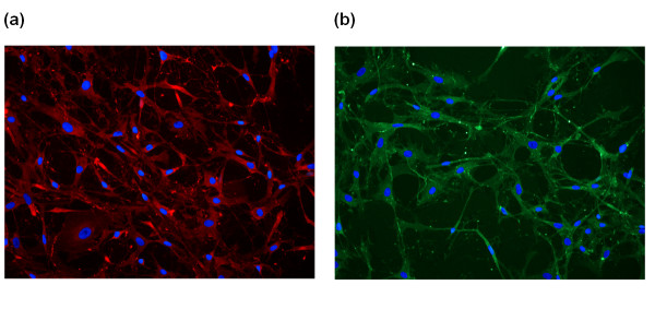
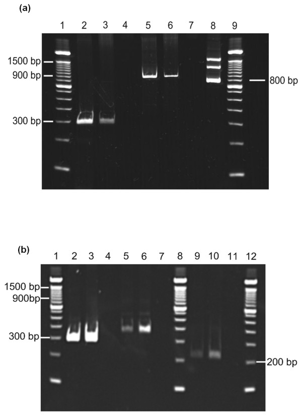
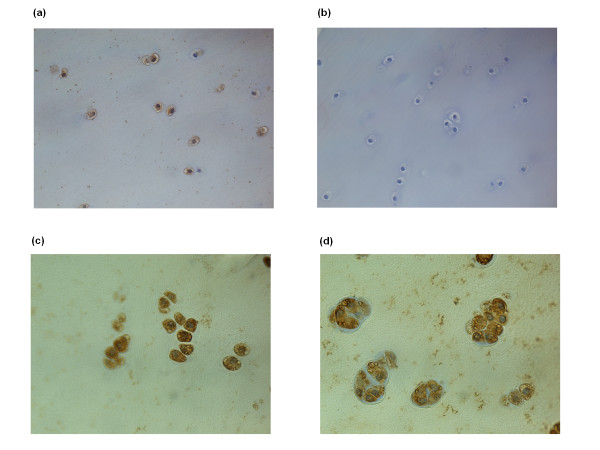
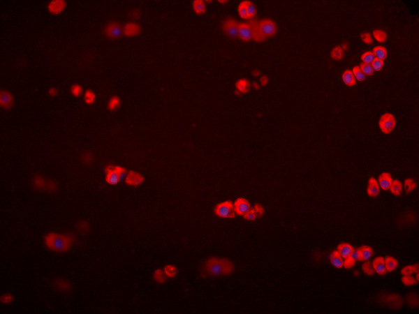
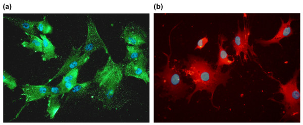
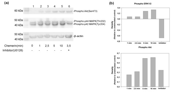
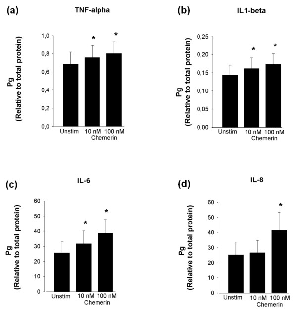
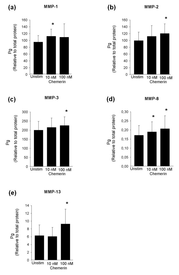
Methods: Tissue sections were taken from human knee joints and labelled with antibodies towards chemerin and ChemR23. Chondrocytes from cartilage tissue were isolated, cultured and assessed for chemerin and ChemR23 expression by PCR and immunolabelling. Receptor activation and intracellular signalling were studied by assessment of phosphorylated mitogen activated protein kinases (MAPKs) and phosphorylated Akt after stimulating cells with recombinant chemerin(21-157). Biological effects of chemerin(21-157) were investigated by measuring secretion of pro-inflammatory cytokines and metalloproteases in cell supernatants.

Results: Both serially cultured human articular chondrocytes and resident cells in native cartilage expressed chemerin and ChemR23. Stimulating cells with chemerin(21-157) resulted in phosphorylation of p44/p42 MAPKs (ERK 1/2) and Akt (Ser 473). Also, significantly enhanced levels of the pro-inflammatory cytokines interleukin-6 (IL-6), interleukin-8 (IL-8), tumour necrosis factor alpha (TNF-α), interleukin-1 beta (IL-1β), and the matrix metalloproteases MMP-1, MMP-2, MMP-3, MMP-8 and MMP-13 were detected.

Conclusions: These results demonstrate that human chondrocytes express both the receptor ChemR23 and the ligand chemerin. Chemerin(21-157) stimulation engaged signal-transduction pathways that further promoted inflammatory signalling in chondrocytes, as judged by an enhanced secretion of cytokines and metalloproteases. Taken together, the previously reported chemotaxis and the present findings suggest that the receptor and its ligand may play pivotal roles in joint inflammation.


Figures

Figure 1
Figure 1
Expression of collagen type II and aggrecan in cultured human articular chondrocytes. (a) Cells were labelled with polyclonal rabbit anti-human collagen type II and secondary antibody conjugated with Alexa Fluor 594 (red). (b) Cells were labelled with monoclonal mouse anti-human aggrecan and secondary antibody conjugated with Alexa Fluor 488 (green). The nuclei were visualized by Dapi dye (blue). Isotype controls had no staining (not shown).
Figure 2
Figure 2
Expression of ChemR23 and chemerin in cultured human articular chondrocytes as detected by RT-PCR. (a) Lanes 2 and 3: RT-PCR with APRT primers and cDNA from two individual cell cultures. Lane 4: APRT primers without cDNA. Lanes 5 and 6: ChemR23 primers and cDNA from two individual cell cultures. Lane 7: ChemR23 primers without cDNA. Lane 8: APRT primers and genomic DNA. (b) Lanes 2 and 3: RT-PCR with APRT primers and cDNA from two individual cell cultures. Lane 4: APRT primers without cDNA. Lanes 5 and 6: Prochemerin primer pair 1 and cDNA from two individual cell cultures. Lane 7: Prochemerin primers without cDNA. Lanes 9 and 10: Prochemerin primer pair 2 and cDNA from two individual cell cultures. Lane 11: Prochemerin primers without cDNA.
Figure 3
Figure 3
The presence of ChemR23 in sections of human articular cartilage. (a) Micrograph A (20X) shows positively (brown) stained chondrocytes in tissue from one patient subjected to total knee arthroplasty. (b) Micrograph B shows an isotype control which was negative. (c and d) Micrograph C (40X) and D (60X) shows positively stained chondrocytes from one patient undergoing reconstruction of ligament.
Figure 4
Figure 4
The presence of chemerin in sections of human articular cartilage. The micrograph (40X) shows positively (red) stained chondrocytes in tissue from one patient undergoing ligament repair. Nuclei were visualized by Dapi dye (blue). Negative controls (isotype IgG) had no red staining (not shown).
Figure 5
Figure 5
The presence of ChemR23 and chemerin in cultured human articular chondrocytes. (a) Cells were labelled with polyclonal rabbit anti-human ChemR23 and secondary antibody conjugated with Alexa Fluor 488 (green). (b) Cells were labelled with polyclonal goat anti-human TIG-2 (chemerin) and secondary antibody conjugated with Alexa Fluor 594 (red). Nuclei were visualized by Dapi dye (blue). Isotype controls had no staining (not shown).
Figure 6
Figure 6
Western blot of phosphorylated p44/42 MAPKs (Thr202/Tyr204) and phosphorylated Akt (Ser 473). (a) Cultured chondrocytes were challenged with 10 nM chemerin21-157 for 1, 2.5, 5 and 10 minutes. Lane 1 represents the control where no chemerin was added and Lane 6 represents the sample extract where the MEK 1/2 kinase inhibitor U0126 was added 1 h prior to a 3.5 minutes chemerin21-157 challenge. (b) The density of each band was normalized to β-actin, the graphs shows the increase in density relative to unstimulated control.
Figure 7
Figure 7
Cytokine levels in supernatants from human articular chondrocytes stimulated with recombinant human chemerin21-157. Cell supernatants were assessed for cytokine contents after 24 h of stimulation with 10 nM or 100 nM recombinant chemerin21-157. The levels of TNF-α (a), IL-1β (b), IL-6 (c), and IL-8 (d) are shown as mean ± standard error of the mean. Results are from six separate experiments analyzed in duplicates. Concentration is given relative to amount of protein (μg/ml). *P < 0.05, stimulated versus unstimulated.
Figure 8
Figure 8
MMP levels in supernatants from human articular chondrocytes stimulated with recombinant human chemerin21-157. Cell supernatants were assessed for content of MMPs after 24 h of stimulation with 10 nM or 100 nM recombinant chemerin21-157. The levels of MMP-1 (a), MMP-2 (b), MMP-3 (c), MMP-8 (d), and MMP-13 (e) are shown as mean ± standard error of the mean values. Results are from six separate experiments analyzed in duplicates. Concentration is given relative to amount of protein (μg/ml). *P < 0.05, stimulated versus unstimulated.

Comment in

References

    1. Wittamer V, Bondue B, Guillabert A, Vassart G, Parmentier M, Communi D. Neutrophil-mediated maturation of chemerin: a link between innate and adaptive immunity. J Immunol. 2005;175:487–493. - PubMed
    1. Meder W, Wendland M, Busmann A, Kutzleb C, Spodsberg N, John H, Richter R, Schleuder D, Meyer M, Forssmann WG. Characterization of human circulating TIG2 as a ligand for the orphan receptor ChemR23. FEBS Lett. 2003;555:495–499. doi: 10.1016/S0014-5793(03)01312-7. - DOI - PubMed
    1. Albanesi C, Scarponi C, Pallotta S, Daniele R, Bosisio D, Madonna S, Fortugno P, Gonzalvo-Feo S, Franssen JD, Parmentier M, De Pita O, Girolomoni G, Sozzani S. Chemerin expression marks early psoriatic skin lesions and correlates with plasmacytoid dendritic cell recruitment. J Exp Med. 2009;206:249–258. doi: 10.1084/jem.20080129. - DOI - PMC - PubMed
    1. Huss RS, Huddleston JI, Goodman SB, Butcher EC, Zabel BA. Synovial tissue-infiltrating natural killer cells in osteoarthritis and peri-prosthetic inflammation. Arthritis Rheum. 2010;62:3799–3805. doi: 10.1002/art.27751. - DOI - PMC - PubMed
    1. Wittamer V, Franssen JD, Vulcano M, Mirjolet JF, Le Poul E, Migeotte I, Brezillon S, Tyldesley R, Blanpain C, Detheux M, Mantovani A, Sozzani S, Vassart G, Parmentier M, Communi D. Specific recruitment of antigen-presenting cells by chemerin, a novel processed ligand from human inflammatory fluids. J Exp Med. 2003;198:977–985. doi: 10.1084/jem.20030382. - DOI - PMC - PubMed

MeSH terms