. 2011 Mar 7;366(1-2):8-19.
doi: 10.1016/j.jim.2010.12.016. Epub 2010 Dec 27.

A robust, high-throughput assay to determine the phagocytic activity of clinical antibody samples

Affiliations

A robust, high-throughput assay to determine the phagocytic activity of clinical antibody samples

Margaret E Ackerman et al. J Immunol Methods. .

Erratum in

  • J Immunol Methods. 2012 Feb 28;376(1-2):156

Abstract

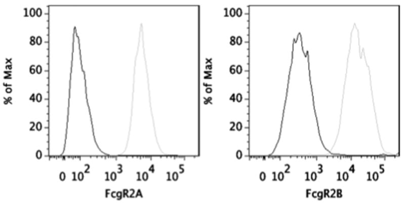
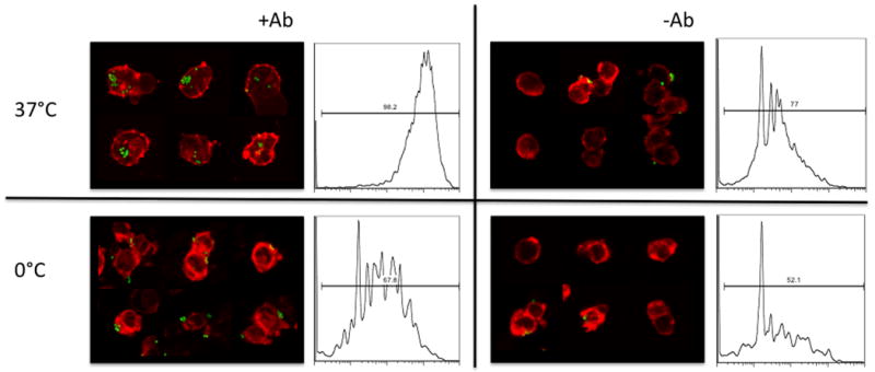
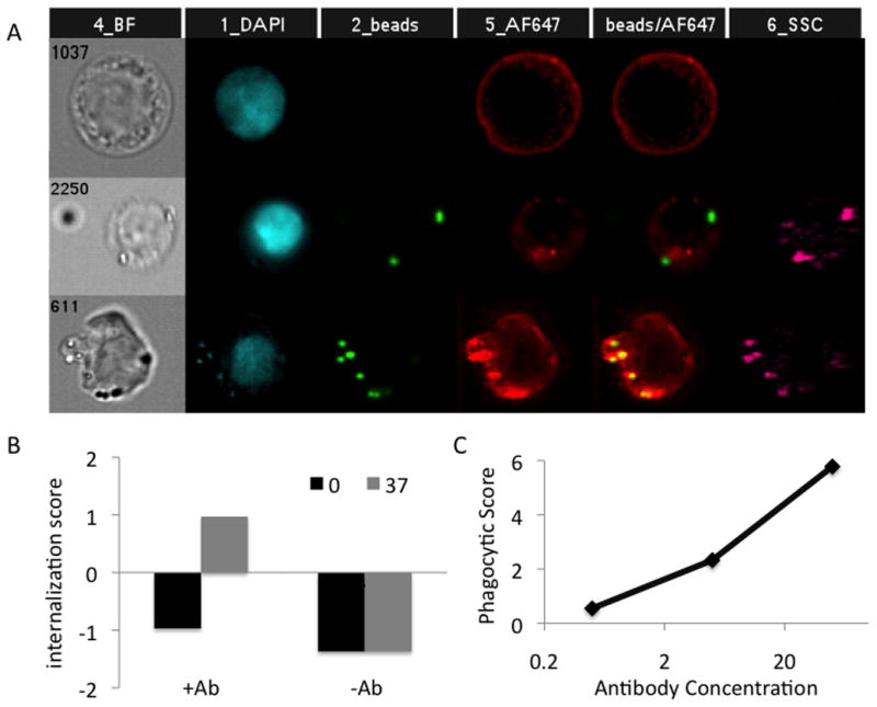
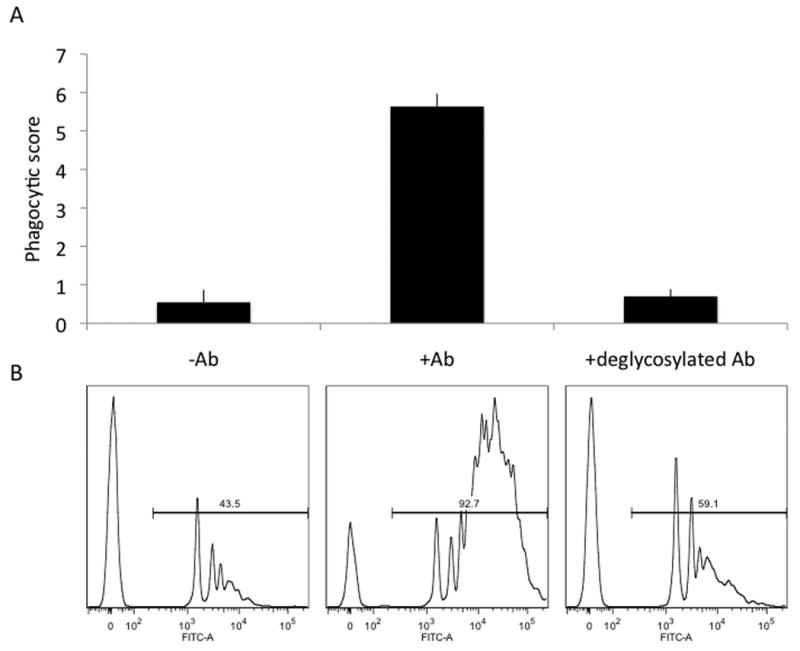
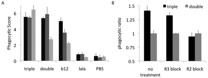
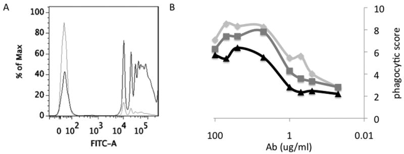
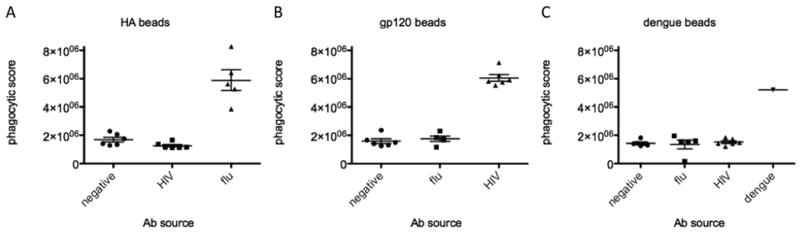
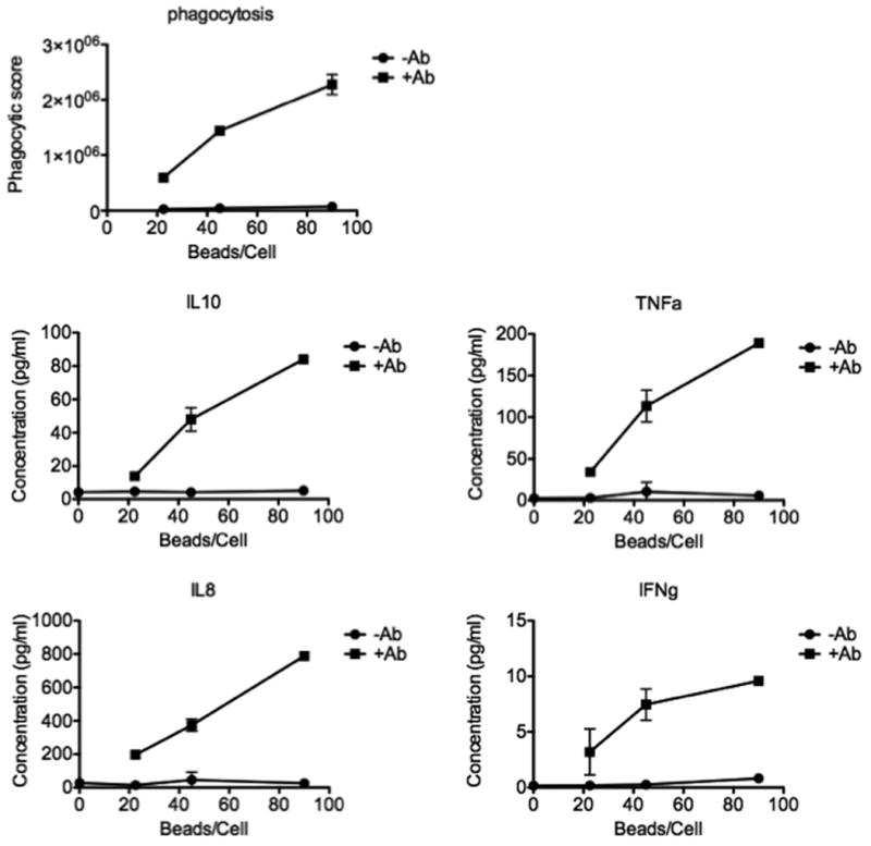
Phagocytosis can be induced via the engagement of Fcγ receptors by antibody-opsonized material. Furthermore, the efficiency of antibody-induced effector functions has been shown to be dramatically modulated by changes in antibody glycosylation. Because infection can modulate antibody glycans, which in turn modulate antibody functions, assays capable of determining the induction of effector functions rather than neutralization or titer provide a valuable opportunity to more fully characterize the quality of the adaptive immune response. Here we describe a robust and high-throughput flow cytometric assay to define the phagocytic activity of antigen-specific antibodies from clinical samples. This assay employs a monocytic cell line that expresses numerous Fc receptors: including inhibitory and activating, and high and low affinity receptors--allowing complex phenotypes to be studied. We demonstrate the adaptability of this high-throughput, flow-based assay to measure antigen-specific antibody-mediated phagocytosis against an array of viruses, including influenza, HIV, and dengue. The phagocytosis assay format further allows for simultaneous analysis of cytokine release, as well as determination of the role of specific Fcγ-receptor subtypes, making it a highly useful system for parsing differences in the ability of clinical and vaccine induced antibody samples to recruit this critical effector function.


Figures

Figure 1
Figure 1. Expression of FcγR2A and 2B
THP-1 cells were labeled with antibodies specific for the extracellular domain of FcγR2A, the Fc receptor implicated in phagocytosis, and the intracellular domain of the inhibitory receptor FcγR2B. Cells labeled with secondary only are traced in black, cells labeled with primary and secondary antibody are traced in gray.
Figure 2
Figure 2. Phagocytosis Assay
Confocal microscopy images and flow cytometry histograms of phagocytosis of green fluorescent beads with or without antibody coating, at 37°C or 0°C. For confocal microscopy, membrane was stained red with the membrane dye, DiI. Histograms represent the fluorescent signal from cell-associated beads. Bead uptake is maximal under conditions where FcγR-mediated phagocytosis can occur.
Figure 3
Figure 3. Confirmation of phagocytosis
A. Representative images captured by the Amnis ImageStreamX Flow Cytometer of cells without any beads (top), surface bound beads (middle), or internalized beads (bottom). Nuclei are stained blue, beads green, and FcγR2 in red. B. Internalization score calculated by Amnis' IDEAS software, indicating internalization of beads when coated by antibody at 37°C, and minimal internalization of beads without antibody-coating, or when incubated at 0°C. C. Dependence of phagocytic score on the density of antibody coating on the surface of the beads.
Figure 4
Figure 4. Dependence on antibody glycosylation state
A. Phagocytic scores and B. Flow cytometry histograms depicting cells incubated with uncoated (-Ab), antibody-coated (+Ab), and deglycosylated antibody-coated (+deglycosylated Ab) fluorescent beads. Uptake of antibody-coated beads is highly dependent on antibody glycosylation state.
Figure 5
Figure 5. Dependence on specific FcγR
A. gp120-coated beads were incubated with b12 variants triple, double, standard and lala (Fc binding knockout) at varying concentrations (100, 20, 5 ng/ml), and the phagocytic score was determined. B. Receptor blocking experiments in which either FcγR2 or FcγR3 was blocked by preincubation with an anti- FcγR antibody. Results are presented as the ratio of the phagocytic score for the variant:double mutant at each given condition.
Figure 6
Figure 6. Antigen specificity of antibody-dependent phagocytosis
A. FACS histogram showing representative data for flu antibodies incubated with dengue beads (gray) or hemagglutinin beads (black). B. Antibody dose-response curve from a single replicate for 3 flu patients are depicted.
Figure 7
Figure 7. Antibody-dependent phagocytosis of patient samples
A. The dot plot represents maximum phagocytic scores of antibodies from negative, HIV positive, or flu positive individuals against hemagglutinin-coated beads. B. Diagrams show phagocytic scores of antibodies from negative, HIV positive, or flu positive individuals against gp120-coated beads. C. Finally, the figure demonstrates phagocytic scores of antibodies from negative, HIV positive, flu positive individuals, or a mouse IgG2A anti-dengue monoclonal against dengue antigen-coated beads.
Figure 8
Figure 8. Cytokine release
Line graphs represent multiplexed analysis of phagocytic scores and corresponding cytokine secretion from cells left untreated, incubated with uncoated beads (circles) or antibody-coated beads (squares) showing strong antibody and dose-dependent phagocytosis-induced release of several cytokines
Figure 9
Figure 9. Time-lapse microscopy of phagocytosis
Antibody-coated (green) and bare (red) beads were incubated with THP-1 cells on coverslips for time lapse microscopy, demonstrating antibody-specific uptake. A. 20× magnification still images representing the first (left) and final (right) frames from two 14 hour experiments. B. Representative 63× magnification images at 14 hours.

References

    1. Allhorn M, Olin AI, Nimmerjahn F, Collin M. Human IgG/Fc gamma R interactions are modulated by streptococcal IgG glycan hydrolysis. PLoS One. 2008;3:e1413. - PMC - PubMed
    1. Anthony RM, Ravetch JV. A novel role for the IgG Fc glycan: the anti-inflammatory activity of sialylated IgG Fcs. J Clin Immunol. 2010;30 1:S9–14. - PubMed
    1. Aslam A, Quinn P, McIntosh RS, Shi J, Ghumra A, McKerrow JH, Bunting KA, Dunne DW, Doenhoff MJ, Morrison SL, Zhang K, Pleass RJ. Proteases from Schistosoma mansoni cercariae cleave IgE at solvent exposed interdomain regions. Mol Immunol. 2008;45:567–74. - PubMed
    1. Berasain P, Carmona C, Frangione B, Dalton JP, Goni F. Fasciola hepatica: parasite-secreted proteinases degrade all human IgG subclasses: determination of the specific cleavage sites and identification of the immunoglobulin fragments produced. Exp Parasitol. 2000;94:99–110. - PubMed
    1. Bernard J, Reveil B, Najman I, Liautaud-Roger F, Fouchard M, Picard O, Cattan A, Mabondzo A, Laverne S, Gallo RC, et al. Discriminating between protective and enhancing HIV antibodies. AIDS Res Hum Retroviruses. 1990;6:243–9. - PubMed

Publication types

MeSH terms

LinkOut - more resources