. 2011 Feb 25;406(3):443-53.
doi: 10.1016/j.jmb.2010.12.025. Epub 2010 Dec 28.

Binding of the inhibitor protein IF(1) to bovine F(1)-ATPase

Affiliations

Binding of the inhibitor protein IF(1) to bovine F(1)-ATPase

John V Bason et al. J Mol Biol. .

Abstract

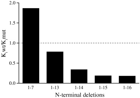
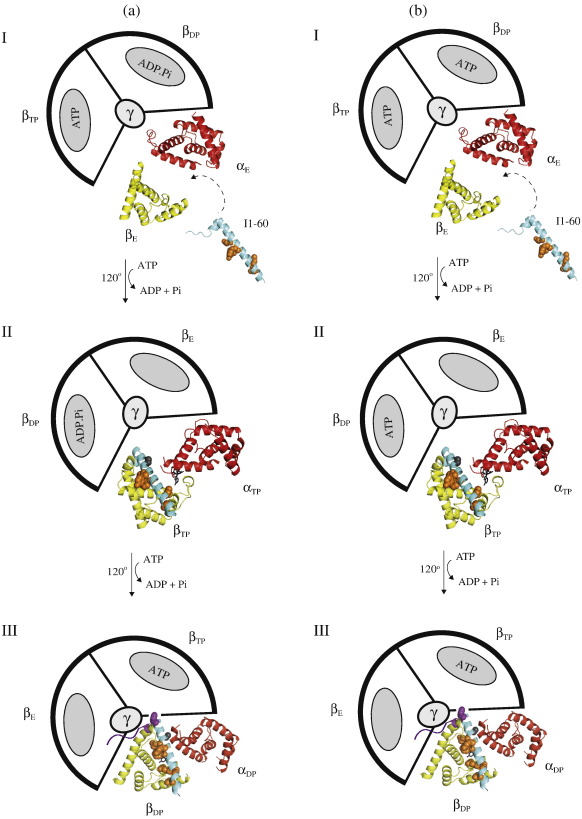
In the structure of bovine F(1)-ATPase inhibited with residues 1-60 of the bovine inhibitor protein IF(1), the α-helical inhibitor interacts with five of the nine subunits of F(1)-ATPase. In order to understand the contributions of individual amino acid residues to this complex binding mode, N-terminal deletions and point mutations have been introduced, and the binding properties of each mutant inhibitor protein have been examined. The N-terminal region of IF(1) destabilizes the interaction of the inhibitor with F(1)-ATPase and may assist in removing the inhibitor from its binding site when F(1)F(o)-ATPase is making ATP. Binding energy is provided by hydrophobic interactions between residues in the long α-helix of IF(1) and the C-terminal domains of the β(DP)-subunit and β(TP)-subunit and a salt bridge between residue E30 in the inhibitor and residue R408 in the C-terminal domain of the β(DP)-subunit. Several conserved charged amino acids in the long α-helix of IF(1) are also required for establishing inhibitory activity, but in the final inhibited state, they are not in contact with F(1)-ATPase and occupy aqueous cavities in F(1)-ATPase. They probably participate in the pathway from the initial interaction of the inhibitor and the enzyme to the final inhibited complex observed in the structure, in which two molecules of ATP are hydrolysed and the rotor of the enzyme turns through two 120° steps. These findings contribute to the fundamental understanding of how the inhibitor functions and to the design of new inhibitors for the systematic analysis of the catalytic cycle of the enzyme.


Figures

None
Graphical abstract
Fig. 1
Fig. 1
The structure of bovine F1-ATPase inhibited with residues 1–60 of the bovine inhibitor protein IF1. The α-, β-, γ-, δ-, and ɛ-subunits are shown in ribbon representation in red, yellow, dark blue, magenta, and green, respectively. The inhibitor protein is light blue. (a) Side view of the complex towards the catalytic interface between the αDP- and βDP-subunits where the inhibitor protein is bound; (b) cross-sectional view of the C-terminal domains of the α- and β-subunits looking up along the axis of the γ-subunit showing interactions of the inhibitor protein with the subunits of F1-ATPase.
Fig. 2
Fig. 2
Alignment of the sequence of residues 1–60 of bovine IF1 with equivalent portions of F1-ATPase inhibitor proteins from other species. The purple, yellow, and green stripes denote strictly conserved, highly conserved, and poorly conserved residues, respectively. The black lines above the sequences mark α-helical regions in bovine IF1. The Expasy accession numbers for inhibitor proteins from Bos taurus, Homo sapiens, Rattus norvegicus, Mus musculus, Saccharomyces cerevisiae, and Pichia jadinii are P01097, Q90112, Q03344, Q35143, P01097, and P09940, respectively.
Fig. 3
Fig. 3
Influence of N-terminal deletions on the binding of the inhibitor protein I1-60GFPHis to bovine F1-ATPase. The data are taken from Supplementary Table S4. For an explanation of the quotient Kimut:Kiwt, see the text.
Fig. 4
Fig. 4
Influence of point mutations in the long α-helix of the inhibitor protein I1-60GFPHis on its binding to bovine F1-ATPase. The mutations and their quantitative effects on binding are given in Supplementary Table S4. Kiwt and Kimut are the values for wild-type and mutant proteins, respectively. Strictly conserved, highly conserved, poorly conserved, and unconserved residues are shown in purple, yellow, green, and white, respectively. The unconserved residues G23, A36, and A38 were not mutated.
Fig. 5
Fig. 5
Residues in the long α-helix of the bovine inhibitor protein that contribute to its binding by interacting directly with subunits of bovine F1-ATPase. The inhibitor protein from residues 8–50 is pale blue, and α-helical regions (residues 14–18 and 21–50) are shown in ribbon representation. The extended N-terminal region (residues 8–13) and the shorter α-helix snake around two antiparallel α-helical regions (residues 1–23 and 232–251) in the γ-subunit, which is part of the central stalk or rotor of the enzyme. Residues with yellow side chains interact with the βDP-subunit; the residue with a red side chain interacts with the αDP-subunit; and the residue with a blue side chain interacts with the βTP-subunit.
Fig. 6
Fig. 6
Charged residues in the long α-helix of the bovine inhibitor protein that are required for the binding of the inhibitor to F1-ATPase but do not interact with F1-ATPase in the structure of the inhibited complex. The long α-helix of IF1 (residues 21–50; light blue) occupies an aqueous cleft between the C-terminal domains of the βDP- and αDP-subunits (yellow and red, respectively). The side chains of amino acids that do not interact with the βDP- and αDP-subunits, but nonetheless are required for the formation of the inhibited complex, are dark blue.
Fig. 7
Fig. 7
Possible schemes of the inhibition of F1-ATPase by the inhibitor protein I1-60. F1-ATPase is depicted as viewed away from the inner mitochondrial membrane from the foot of the central stalk. For simplicity, only those α- and β-subunits that form the catalytic interface, where I1-60 binds, are shown in ribbon representation. The α- and β-subunits are in red and yellow, respectively, and I1-60 is in light blue. The remaining α- and β-subunits and the γ-subunit are represented schematically, and the corresponding α-subunits have been omitted for simplicity. Panel A: In state I, IF1 binds first via the longer α-helix to the C-terminal domain of the βE-subunit in the catalytic interface between the αE- and βE-subunits when the enzyme is in the ground state [PDB code 2JDI]. These initial interactions between inhibitor and enzyme involve many of the group 1 residues, E30, Y33, F34, Q41, L42, and L45 (orange), and the formation of this and subsequent intermediates may also include the group 2 charged residues, R25, Q27, R37, K39, and Q41. In state II, rotation of the γ-subunit through 120° closes the αEβE interface, converting it to the αTPβTP interface [PDB was made by docking I1-60 into the αTPTP interface of the azide free ground state structure (PDB code 2JDI) at the same angle to which it binds to the αDPβDP interface in the final inhibited state (PDB code 2v7q)6]. This αTP βTP interface is similar in structure but not identical to the αDP βDP interface, and many of the interactions observed in the structure of the final inhibited state probably will be present at this stage. They include the interactions formed by the group 1 residues of IF1 (E30, Y33, F34, Q41, L42, and L45; orange). Also, it is possible that the group 1 residue A28 (grey) may bind to the αTP-subunit at this point. However, residue F22 cannot interact with F1-ATPase at this stage. In state III, further rotation of the γ-subunit through 120° leads to the formation of the final inhibited structure [PDB code 2v7q], either, as depicted in scheme A, a dead end state, where an ATP molecule has been hydrolysed at the same catalytic interface that I1-60 binds, or, as shown in scheme B, a pre-hydrolysis state, where an ATP molecule has not been hydrolysed. Residue F22 (purple) can now interact with the βTP-subunit, completing the binding interactions between the long α-helix of IF1 and F1-ATPase. In this final inhibited state, the N-terminal region (purple) will also interact with the γ- and αE-subunits.

References

    1. Pullman M.E., Monroy G.C. A soluble heat stable protein in mitochondria from bovine heart that inhibits ATP hydrolase activity. J. Biol. Chem. 1963;238:3762–3769. - PubMed
    1. Cabezón E., Montgomery M.G., Leslie A.G.W., Walker J.E. The structure of bovine F1-ATPase in complex with its regulatory protein IF1. Nat. Struct. Biol. 2003;10:744–750. - PubMed
    1. Lippe G., Sorgato M.C., Harris D.A. The binding and release of the inhibitor protein are governed independently by ATP and membrane potential in ox-heart submitochondrial vesicles. Biochim. Biophys. Acta. 1988;933:12–21. - PubMed
    1. Walker J.E., Gay N.J., Powell S.J., Kostina M., Dyer M.R. ATP synthase from bovine mitochondria: sequences of imported precursors of oligomycin sensitivity conferral protein, factor 6, and adenosine triphosphatase inhibitor protein. Biochemistry. 1987;26:8613–8619. - PubMed
    1. Cabezón E., Runswick M.J., Leslie A.G.W., Walker J.E. The structure of bovine IF1, the regulatory subunit of mitochondrial F1-ATPase. EMBO. J. 2001;20:6990–6996. - PMC - PubMed

Publication types

LinkOut - more resources