

Brain network connectivity in individuals with schizophrenia and their siblings
- PMID: 21193174
- PMCID: PMC3081915
- DOI: 10.1016/j.biopsych.2010.11.009
Brain network connectivity in individuals with schizophrenia and their siblings
Abstract
Background: Research on brain activity in schizophrenia has shown that changes in the function of any single region cannot explain the range of cognitive and affective impairments in this illness. Rather, neural circuits that support sensory, cognitive, and emotional processes are now being investigated as substrates for cognitive and affective impairments in schizophrenia, a shift in focus consistent with long-standing hypotheses about schizophrenia as a disconnection syndrome. Our goal was to further examine alterations in functional connectivity within and between the default mode network and three cognitive control networks (frontal-parietal, cingulo-opercular, and cerebellar) as a basis for such impairments.
Methods: Resting state functional magnetic resonance imaging was collected from 40 individuals with DSM-IV-TR schizophrenia, 31 siblings of individuals with schizophrenia, 15 healthy control subjects, and 18 siblings of healthy control subjects while they rested quietly with their eyes closed. Connectivity metrics were compared between patients and control subjects for both within- and between-network connections and were used to predict clinical symptoms and cognitive function.
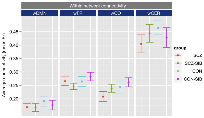
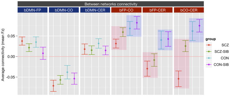
Results: Individuals with schizophrenia showed reduced distal and somewhat enhanced local connectivity between the cognitive control networks compared with control subjects. Additionally, greater connectivity between the frontal-parietal and cerebellar regions was robustly predictive of better cognitive performance across groups and predictive of fewer disorganization symptoms among patients.
Conclusions: These results are consistent with the hypothesis that impairments of executive function and cognitive control result from disruption in the coordination of activity across brain networks and additionally suggest that these might reflect impairments in normal pattern of brain connectivity development.
Copyright © 2011 Society of Biological Psychiatry. Published by Elsevier Inc. All rights reserved.
Figures

References
-
- Van Snellenberg JX, Torres IJ, Thornton AE. Functional neuroimaging of working memory in schizophrenia: task performance as a moderating variable. Neuropsychology. 2006;20:497–510. - PubMed
-
- Achim AM, Lepage M. Episodic memory-related activation in schizophrenia: Meta-analysis. British Journal of Psychiatry. 2005;187:500–509. - PubMed
Publication types
MeSH terms
Substances
Grants and funding
LinkOut - more resources
Full Text Sources
Medical
