

Detection of coronary atherosclerotic plaques with superficial proteoglycans and foam cells using real-time intrinsic fluorescence spectroscopy
- PMID: 21193196
- PMCID: PMC3049853
- DOI: 10.1016/j.atherosclerosis.2010.11.020
Detection of coronary atherosclerotic plaques with superficial proteoglycans and foam cells using real-time intrinsic fluorescence spectroscopy
Abstract
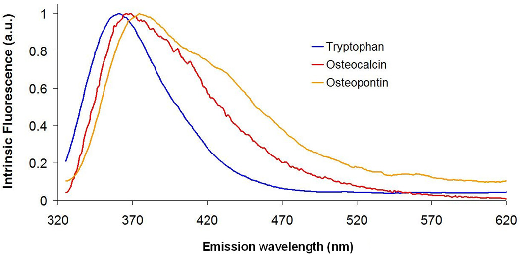
Objectives: The protein components of low-density lipoprotein (LDL), oxidized LDL and proteoglycans such as versican contain tryptophan, an amino acid with characteristic fluorescence features at 308 nm excitation wavelength. We hypothesize that intrinsic fluorescence spectroscopy at 308 nm excitation wavelength IFS308, a method suitable for clinical use, can identify coronary artery lesions with superficial foam cells (SFCs) and/or proteoglycans.
Methods: We subjected 119 human coronary artery specimens to in vitro fluorescence and reflectance spectroscopy. We used 5 basis spectra to model IFS308, and extracted their contributions to each individual IFS308 spectrum. A diagnostic algorithm using the contributions of Total Tryptophan and fibrous cap to IFS308 was built to identify specimens with SFCs and/or proteoglycans in their top 50 μm.
Results: We detected SFCs and/or proteoglycans, such as versican or the glycosaminoglycan hyaluronan, in 24 fibrous cap atheromas or pathologic intimal thickening (PIT) lesions. An algorithm using the contributions of Total Tryptophan and fibrous cap to IFS308 was able to identify these segments with 92% sensitivity and 80% specificity.
Conclusion: We were able to establish a set of characteristic LDL, oxidized LDL, versican and hyaluronan fluorescence spectra, ready to be used for real-time diagnosis. The IFS(308) technique detects SFCs and/or proteoglycans in fibrous cap atheromas and PIT lesions. SFCs and proteoglycans are histological markers of vulnerable plaques, and this study is a step further in developing an invasive clinical tool to detect the vulnerable atherosclerotic plaque.
Copyright © 2010 Elsevier Ireland Ltd. All rights reserved.
Conflict of interest statement
There are no conflicts of interest for any of the authors listed.
Figures


Similar articles
-
Intrinsic fluorescence and diffuse reflectance spectroscopy identify superficial foam cells in coronary plaques prone to erosion.Arterioscler Thromb Vasc Biol. 2006 Jul;26(7):1594-600. doi: 10.1161/01.ATV.0000225699.36212.23. Epub 2006 May 4. Arterioscler Thromb Vasc Biol. 2006. PMID: 16675721
-
Intrinsic versus laser-induced fluorescence spectroscopy for coronary atherosclerosis: a generational comparison model for testing diagnostic accuracy.Appl Spectrosc. 2012 Dec;66(12):1403-10. doi: 10.1366/11-06566. Appl Spectrosc. 2012. PMID: 23231902
-
Differential accumulation of proteoglycans and hyaluronan in culprit lesions: insights into plaque erosion.Arterioscler Thromb Vasc Biol. 2002 Oct 1;22(10):1642-8. doi: 10.1161/01.atv.0000034021.92658.4c. Arterioscler Thromb Vasc Biol. 2002. PMID: 12377743
-
The Initial Human Atherosclerotic Lesion and Lipoprotein Modification-A Deep Connection.Int J Mol Sci. 2021 Oct 25;22(21):11488. doi: 10.3390/ijms222111488. Int J Mol Sci. 2021. PMID: 34768918 Free PMC article. Review.
-
An integrated approach to simulating the vulnerable atherosclerotic plaque.Am J Physiol Heart Circ Physiol. 2020 Oct 1;319(4):H835-H846. doi: 10.1152/ajpheart.00174.2020. Epub 2020 Aug 14. Am J Physiol Heart Circ Physiol. 2020. PMID: 32795179 Free PMC article. Review.
Cited by
-
Non-destructive detection of matrix stabilization correlates with enhanced mechanical properties of self-assembled articular cartilage.J Tissue Eng Regen Med. 2019 Apr;13(4):637-648. doi: 10.1002/term.2824. Epub 2019 Mar 20. J Tissue Eng Regen Med. 2019. PMID: 30770656 Free PMC article.
-
In vivo label-free structural and biochemical imaging of coronary arteries using an integrated ultrasound and multispectral fluorescence lifetime catheter system.Sci Rep. 2017 Aug 21;7(1):8960. doi: 10.1038/s41598-017-08056-0. Sci Rep. 2017. PMID: 28827758 Free PMC article.
-
Current Challenges in Understanding the Cellular and Molecular Mechanisms in Niemann-Pick Disease Type C1.Int J Mol Sci. 2019 Sep 6;20(18):4392. doi: 10.3390/ijms20184392. Int J Mol Sci. 2019. PMID: 31500175 Free PMC article. Review.
-
Detection of glycosaminoglycan loss in articular cartilage by fluorescence lifetime imaging.J Biomed Opt. 2018 Dec;23(12):1-8. doi: 10.1117/1.JBO.23.12.126002. J Biomed Opt. 2018. PMID: 30578627 Free PMC article.
-
The biochemistry and immunohistochemistry of versican.Methods Cell Biol. 2018;143:261-279. doi: 10.1016/bs.mcb.2017.08.015. Epub 2017 Nov 26. Methods Cell Biol. 2018. PMID: 29310782 Free PMC article.
References
-
- Kramer MC, Rittersma SZ, Winter RJ, et al. Relationship of Thrombus Healing to Underlying Plaque Morphology in Sudden Coronary Death. J Am Coll Cardiol. 2010;55:122–132. - PubMed
-
- Farb A, Burke AP, Tang AL, et al. Coronary plaque erosion without rupture into a lipid core. A frequent cause of coronary thrombosis in sudden coronary death. Circulation. 1996;93:1354–1363. - PubMed
-
- Wight TN, Merrilees MJ. Proteoglycans in atherosclerosis and restenosis: key roles for versican. Circ Res. 2004;94:1158–1167. - PubMed
-
- Gutierrez P, O'Brien KD, Ferguson M, Nikkari ST, Alpers CE, Wight TN. Differences in the distribution of versican, decorin, and biglycan in atherosclerotic human coronary arteries. Cardiovasc Pathol. 1997;6:271–278. - PubMed
Publication types
MeSH terms
Substances
Grants and funding
LinkOut - more resources
Full Text Sources
Miscellaneous
