. 2011 Apr;96(4):543-51.
doi: 10.3324/haematol.2010.027557. Epub 2010 Dec 29.

Establishment of a xenograft model of human myelodysplastic syndromes

Affiliations

Establishment of a xenograft model of human myelodysplastic syndromes

Yukari Muguruma et al. Haematologica. 2011 Apr.

Abstract

Background: To understand how myelodysplastic syndrome cells evolve from normal stem cells and gain competitive advantages over normal hematopoiesis, we established a murine xenograft model harboring bone marrow cells from patients with myelodysplastic syndromes or acute myeloid leukemia with myelodysplasia-related changes.

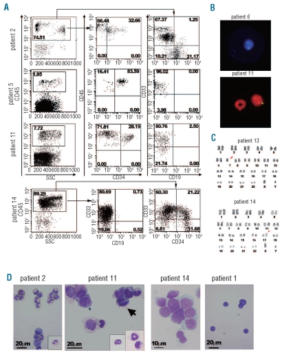
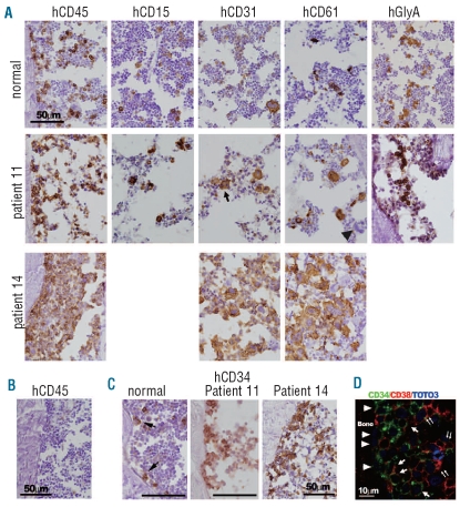
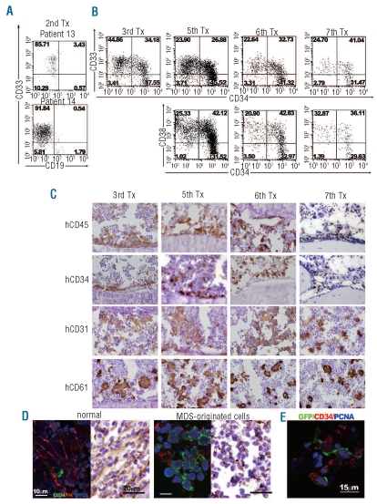
Design and methods: Bone marrow CD34(+) cells obtained from patients were injected, with or without human mesenchymal stem cells, into the bone marrow of non-obese diabetic/severe combined immunodeficient/IL2Rγ(null) hosts. Engraftment and differentiation of cells derived from the patients were investigated by flow cytometry and immunohistochemical analysis.

Results: Co-injection of patients' cells and human mesenchymal stem cells led to successful engraftment of patient-derived cells that maintained the immunophenotypes and genomic abnormalities of the original patients. Myelodysplastic syndrome-originated clones differentiated into mature neutrophils, megakaryocytes, and erythroblasts. Two of the samples derived from patients with acute myeloid leukemia with myelodysplasia-related changes were able to sustain neoplastic growth into the next generation while these cells had limited differentiation ability in the murine host. The hematopoiesis of mice engrafted with patients' cells was significantly suppressed even when human cells accounted for less than 1% of total marrow mononuclear cells. Histological studies revealed invasion of the endosteal surface by patient-derived CD34(+) cells and disruption of extracellular matrix architecture, which probably caused inhibition of murine hematopoiesis.

Conclusions: We established murine models of human myelodysplastic syndromes using cells obtained from patients: the presence of neoplastic cells was associated with the suppression of normal host hematopoiesis. The efficiency of engraftment was related to the presence of an abnormality in chromosome 7.


Figures

Figure 1.
Figure 1.
Engraftment of human MDS-originated hematopoietic cells in the bone marrow (BM) of NOG mice. (A) Representative flow cytometric profiles of BM cells recovered from mice engrafted with patients’ BM cells. The majority of human CD45-expressing cells were positive for a myeloid marker CD33 in patients 5, 11, and 14, while some CD19+ cells were present in BM cells recovered from the mouse engrafted with cells from patient 2. For patient 14, approximately one quarter of CD33+ cells co-expressed CD34. The percentages of cells in the respective regions are shown. (B) FISH detection of a partial deletion of chromosome 7 and monosomy 7. Human cells recovered from the mice engrafted with BM cells from patient 6 and patient 11 were subjected to FISH analysis using D7Z1 (green signal for centromere of chromosome 7) plus D7S486 (red signal for 7q31 region) probes for patient 6 and D7Z1 (yellow signal) probe for patient 11. In a lower panel, a murine granulocyte with a ring-shaped nucleus which did not hybridize with the human probe is located adjacent to the human cell hybridized with D721. All cells analyzed (10 cells for patient 6 and 100 cells for patient 11) demonstrated the same outcome. (C) Chromosomal analysis of cells recovered from the mice transplanted with MDS-originated cells obtained from the BM of patient 13 and patient 14 demonstrated the maintenance of the original abnormal karyotype, namely isochromosome 17 and monosomy 7 (arrows), respectively. Eight cells were analyzed for patient 13 and 20 cells for patient 14. (D) Wright-Giemsa-stained cytospin preparations made of CD45-sorted human cells. In the cytospin samples for patient 2, various stages of myeloid lineage cells and an eosinophil are shown. An insert shows a myelocyte with pseudo-Pelger anomaly. For patient 11, an arrow indicates a bi-nucleated myelocyte. Inserts show differentiated neutrophils. The majority of cells found in a cytospin preparation of BM cells obtained from the mice engrafted with cells from patient 14 demonstrated fine chromatin formation and conspicuous nucleoli. Cytospin samples of a normal cell-engrafted mouse (patient 1) were composed of lymphocytes.
Figure 2.
Figure 2.
Histological analysis of human MDS-originated cells in murine bone marrow (BM). (A) Immunohistochemical staining of bone sections of the mice engrafted with BM cells from normal individuals and patients 11 and 14. Human cells were recognized by specific staining for human antigens. Cells that reacted with antibodies specific to human CD45, CD15, CD31, CD61, and glycophorin A (GlyA) were detected throughout the murine BM compartment in the bone samples of normal individuals and patient 11. Relatively small megakaryocytes with separated nuclei (an arrow) were often observed in the CD31 stained sections of the mice engrafted with cells from patient 11. Note, murine megakaryocytes are negative for human CD61 (an arrowhead), confirming the specificity of the antigen-antibody reaction. In bone samples of patient 14, human CD45-expressing cells occupied most of the marrow compartment. Cells expressing megakaryocytic markers, CD31 and CD61, were noticeable. (B) A human CD45-stained bone section of contra-lateral tibia of the mice intramedullary injected with MDS-originated bone marrow CD34+ cells from patient 11 and MSC. Neither obvious hypocellularity nor human CD45+ cells were detected. (C) In bone samples of patient 11, MDS-originated CD34+ cells proliferated along the surface of the endosteum, while individual CD34+ cells (arrows) attached to the endosteum in normal cell-engrafted mice. Invasion of CD34+ cells was prominent in bones engrafted with cells from patient 14. (D) The cells expressing only CD34 (arrowheads) attached to the endosteum, but the cells expressing both CD34 and CD38 did not (arrows). The cells expressing only CD38 (double arrows) were located distant from the bone.
Figure 3.
Figure 3.
Serial transplantation of MDS-originated cells and the bone marrow microenvironment (A) Flow cytometric profiles of bone marrow (BM) cells recovered from secondary hosts. Tx indicates transplantation. (B) Flow cytometric profiles of BM cells recovered from mice that had undergone serial transplants with BM cells from patient 14. (C) Immunohistochemical staining of bones obtained from mice serially transplanted with cells of patient 14 demonstrated a lineage cell staining pattern similar to that of the primary recipient mice, while CD45 and CD34-expressing cells were more confined to the endosteal region. (D) Immunofluorescent and immunohisotochemical staining for fibronectin (FN) of bones of normal cell- and MDS-originated cell-engrafted mice. Murine and human cells in normal cell-transplanted mice were tightly enveloped by fibronectin while the fibronectin network of MDS-originated cell-engrafted mice was disrupted. A light field photograph confirmed the well-structured fibronectin network in the BM of normal cell-engrafted mice, but only fibronectin fibrils were detected in the MDS-originated cell-engrafted mice. Stained sections of the mice engrafted with cells from patient 11 are shown. The same staining patterns were confirmed in bone sections of mice engrafted with cells from patients 13 and 14. (E) MDS-originated CD34+ cells expressing a proliferating marker, PCNA, interacted with human MSC marked with green fluorescent protein.

References

    1. Steensma DP, Tefferi A. The myelodysplastic syndrome(s): a perspective and review highlighting current controversies. Leuk Res. 2003;27(2):95–120. - PubMed
    1. Nimer SD. Myelodysplastic syndromes. Blood. 2008;111 (10):4841–51. - PubMed
    1. Heaney ML, Golde DW. Myelodysplasia. N Engl J Med. 1999;340(21):1649–60. - PubMed
    1. Haase D, Fonatsch C, Freund M, Wormann B, Bodenstein H, Bartels H, et al. Cytogenetic findings in 179 patients with myelodysplastic syndromes. Ann Hematol. 1995;70(4):171–87. - PubMed
    1. Parlier V, van Melle G, Beris P, Schmidt PM, Tobler A, Haller E, et al. Hematologic, clinical, and cytogenetic analysis in 109 patients with primary myelodysplastic syndrome. Prognostic significance of morphology and chromosome findings. Cancer Genet Cytogenet. 1994;78(2):219–31. - PubMed

Publication types