. 2011 Feb;134(Pt 2):449-63.
doi: 10.1093/brain/awq347. Epub 2010 Dec 29.

White matter damage and cognitive impairment after traumatic brain injury

Affiliations

White matter damage and cognitive impairment after traumatic brain injury

Kirsi Maria Kinnunen et al. Brain. 2011 Feb.

Abstract

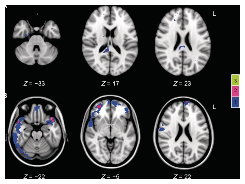
White matter disruption is an important determinant of cognitive impairment after brain injury, but conventional neuroimaging underestimates its extent. In contrast, diffusion tensor imaging provides a validated and sensitive way of identifying the impact of axonal injury. The relationship between cognitive impairment after traumatic brain injury and white matter damage is likely to be complex. We applied a flexible technique-tract-based spatial statistics-to explore whether damage to specific white matter tracts is associated with particular patterns of cognitive impairment. The commonly affected domains of memory, executive function and information processing speed were investigated in 28 patients in the post-acute/chronic phase following traumatic brain injury and in 26 age-matched controls. Analysis of fractional anisotropy and diffusivity maps revealed widespread differences in white matter integrity between the groups. Patients showed large areas of reduced fractional anisotropy, as well as increased mean and axial diffusivities, compared with controls, despite the small amounts of cortical and white matter damage visible on standard imaging. A stratified analysis based on the presence or absence of microbleeds (a marker of diffuse axonal injury) revealed diffusion tensor imaging to be more sensitive than gradient-echo imaging to white matter damage. The location of white matter abnormality predicted cognitive function to some extent. The structure of the fornices was correlated with associative learning and memory across both patient and control groups, whilst the structure of frontal lobe connections showed relationships with executive function that differed in the two groups. These results highlight the complexity of the relationships between white matter structure and cognition. Although widespread and, sometimes, chronic abnormalities of white matter are identifiable following traumatic brain injury, the impact of these changes on cognitive function is likely to depend on damage to key pathways that link nodes in the distributed brain networks supporting high-level cognitive functions.


Figures

Figure 1
Figure 1
Lesion probability maps of (A) white matter lesions visible on gradient echo imaging and (B) contusions. The colour bar indicates the number of patients who had lesions at each site. Green–yellow indicates where lesions were present in three (11%) of the 28 patients with traumatic brain injury, pink indicates where they were present in two (7%) and blue where a lesion was found in one patient only.
Figure 2
Figure 2
Widespread white matter disruption following traumatic brain injury. Axial slices of the results of (A) fractional anisotropy (FA), (B) mean diffusivity (MD), (C) axial diffusivity (Dax) and (D) radial diffusivity (Drad) TBSS contrasts between traumatic brain injury and control groups. Fractional anisotropy (red): controls > traumatic brain injury; mean diffusivity (dark blue): traumatic brain injury > controls; Dax (yellow): traumatic brain injury > controls; and Drad (light blue): traumatic brain injury > controls. The contrasts are overlaid on a standard Montréal Neurological Institute 152 T1 1 mm brain and the mean fractional anisotropy skeleton (in green) with display thresholds set to range from 0.2 to 0.8. The results are thresholded at P ≤ 0.05, corrected for multiple comparisons.
Figure 3
Figure 3
Patients with microbleed evidence of diffuse axonal injury show more extensive white matter damage. The results of (A) fractional anisotropy (FA), (B) mean diffusivity (MD) and (C) radial diffusivity (Drad) TBSS contrasts between patient groups with and without microbleed evidence of diffuse axonal injury. Fractional anisotropy (red): non-microbleed > microbleed; mean diffusivity (dark blue): microbleed > non-microbleed; and Drad (light blue): microbleed > non-microbleed. The contrasts are overlaid on a standard Montréal Neurological Institute 152 T1 1 mm brain and the mean fractional anisotropy skeleton (in green). The results are thresholded at P ≤ 0.05, corrected for multiple comparisons.
Figure 4
Figure 4
Patients without microbleeds also show evidence of white matter damage. The results of (A) fractional anisotropy (FA), (B) mean diffusivity (MD) and (C) axial diffusivity (Dax) TBSS contrasts between patients without microbleed evidence of diffuse axonal injury (non-microbleed) and controls. Fractional anisotropy (red): controls > non-microbleed, mean diffusivity (dark blue): non-microbleed > controls; and Dax (yellow): non-microbleed > controls. The contrasts are overlaid on a standard Montréal Neurological Institute 152 T1 1 mm brain and the mean fractional anisotropy skeleton (in green). The results are thresholded at P ≤ 0.05, corrected for multiple comparisons.
Figure 5
Figure 5
The results of TBSS regression analysis of associative learning and memory (People Test immediate recall total) by fractional anisotropy (FA) across the traumatic brain injury and control groups. (A) Areas where fractional anisotropy is positively correlated with People Test (PT) recall score across the two groups are indicated in red (FA/PT: ALL+). The result is overlaid on a standard Montréal Neurological Institute 152 T1 1 mm brain and the mean fractional anisotropy skeleton (in green). For display purposes the result is displayed with a multiple comparisons threshold of P ≤ 0.1. (B) Graph showing individual data points in both groups for People Test recall score against fractional anisotropy in the peak voxel (Montréal Neurological Institute x = 7, y = −5, z = 9). A second-order polynomial regression slope is shown, which provides a more accurate fit than the linear regression identified by the whole-brain general linear model analysis. CON = control; TBI = traumatic brain injury.
Figure 6
Figure 6
The results of TBSS regression analysis of the group interaction between alternating-switch cost (Trail Making Test Trails B minus Trail A) and radial diffusivity (Drad) in the traumatic brain injury and control groups. (A) Results of the whole-brain analysis with significant areas of the interaction effect for Drad (TBI+/CON−) shown in light blue. The results are thresholded at P ≤ 0.01, corrected for multiple comparisons and overlaid on a standard Montréal Neurological Institute 152 T1 1 mm brain and the mean fractional anisotropy skeleton (in green). (B) Graph illustrating linear regression slopes for each group and individual data points for alternating-switch cost against Drad in the peak voxel (Montréal Neurological Institute x = 18, y = −38, z = 36) of the interaction effect. Drad values are expressed as mm2/s × 10−3 for convenience of display. CON = control; TBI = traumatic brain injury.

References

    1. Aggleton JP EPS Mid-Career Award 2006. Understanding anterograde amnesia: disconnections and hidden lesions (Review) Q J Exp Psychol. 2008;61:1441–71. - PubMed
    1. Aggleton JP, Brown MW. Episodic memory, amnesia, and the hippocampal–anterior thalamic axis. Behav Brain Sci. 1999;22:425–89. - PubMed
    1. Arfanakis K, Haughton VM, Carew JD, Rogers BP, Dempsey RJ, Meyerand ME. Diffusion tensor MR imaging in diffuse axonal injury. Am J Neuroradiol. 2002;23:794–802. - PMC - PubMed
    1. Assaf Y, Pasternak O. Diffusion tensor imaging (DTI)-based white matter mapping in brain research: A review (Review) J Mol Neurosci. 2008;34:51–61. - PubMed
    1. Baddeley AD, Emslie H, Nimmo-Smith I. Bury-St-Edmunds: Thames Valley Test Company; 1994. Doors and people test: a test of visual and verbal recall and recognition.

Publication types

MeSH terms