. 2011 Jan 18;108(3):1040-5.
doi: 10.1073/pnas.0914906108. Epub 2010 Dec 30.

Possible roles for Munc18-1 domain 3a and Syntaxin1 N-peptide and C-terminal anchor in SNARE complex formation

Affiliations

Possible roles for Munc18-1 domain 3a and Syntaxin1 N-peptide and C-terminal anchor in SNARE complex formation

Shu-Hong Hu et al. Proc Natl Acad Sci U S A. .

Abstract

Munc18-1 and Syntaxin1 are essential proteins for SNARE-mediated neurotransmission. Munc18-1 participates in synaptic vesicle fusion via dual roles: as a docking/chaperone protein by binding closed Syntaxin1, and as a fusion protein that binds SNARE complexes in a Syntaxin1 N-peptide dependent manner. The two roles are associated with a closed-open Syntaxin1 conformational transition. Here, we show that Syntaxin N-peptide binding to Munc18-1 is not highly selective, suggesting that other parts of the SNARE complex are involved in binding to Munc18-1. We also find that Syntaxin1, with an N peptide and a physically anchored C terminus, binds to Munc18-1 and that this complex can participate in SNARE complex formation. We report a Munc18-1-N-peptide crystal structure that, together with other data, reveals how Munc18-1 might transit from a conformation that binds closed Syntaxin1 to one that may be compatible with binding open Syntaxin1 and SNARE complexes. Our results suggest the possibility that structural transitions occur in both Munc18-1 and Syntaxin1 during their binary interaction. We hypothesize that Munc18-1 domain 3a undergoes a conformational change that may allow coiled-coil interactions with SNARE complexes.


Conflict of interest statement

The authors declare no conflict of interest.

Figures
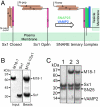
Fig. 1.
Fig. 1.
Munc18-1 interaction with Sx1. (A) Sx1 Conformations. Sx1 is a helical protein comprising an N peptide (residues 1–10; labeled N-pep), a 3-helical N-terminal domain termed Habc (helices labeled A, B, and C) linked to a SNARE helix H3 (labeled 3) that is tethered to the plasma membrane at the C terminus by a transmembrane region. In the closed Sx1 conformation, the H3 helix interacts with the Habc domain and is unavailable to interact with SNARE partner proteins SNAP25 and VAMP2 (also called Synaptobrevin). When Sx1 adopts an open conformation, the H3 SNARE motif is available to interact with SNARE partner proteins SNAP25 (two helices, green, attached to the plasma membrane via cysteine palmitoylation) and VAMP2 (blue, tethered to the vesicle membrane by a transmembrane region at its C terminus) to form the SNARE ternary complex in a process that drives vesicle fusion. Munc18-1 is known to interact with the closed conformation of Sx1 and with the SNARE ternary complex; the latter interaction is dependent on the Sx1 N peptide. (B) Sx1-His–Munc18-1. Sx1-His immobilized on resin forms a complex with Munc18-1. (C) Sx1 bound to Munc18-1 participates in SNARE complex formation. Sx1 immobilized on resin by a C-terminal His tag was used alone or in complex with Munc18-1 (B) to investigate the formation of SNARE complex. Lane 1 shows the formation of SNARE ternary complex (Sx1/SNAP25/VAMP2) in the absence of Munc18-1; lane 2 shows that preformed Munc18-1–Sx1 complex also interacts with SNAP25 and VAMP2. For comparison, lane 3 shows the relative amounts of these proteins when Munc18-1 is incubated with preformed SNARE complex.
Fig. 2.
Fig. 2.
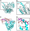
Structure of Munc18-1 complexed with the Sx4 N peptide. (A) Ribbon diagram of the structure of Munc18-1 (cyan) bound to the Sx4 N peptide (Sx41–10) (orange). Domains 1, 2, and 3 of Munc18-1 are labeled D1, D2, and D3, respectively. (B) Overlay of domains 2 and 3 of the two molecules of Munc18-1 in the asymmetric unit of the crystal structure (cyan, yellow), showing that domain 1 can rotate with respect to domains 2 and 3. (C. and D) Ribbon diagrams showing that the monomer–monomer interface observed in the crystal structure of (C) Munc18-1–Sx4 N peptide (cyan, yellow) resembles that in the crystal structure of (D) Munc18-3–Sx4 N peptide (green, purple). Bound Sx4 N peptides are shown in orange space-filling representation.
Fig. 3.
Fig. 3.
Munc18-1 adopts a conformation that may preclude closed Sx1. (A) Superposition of Munc18-1 structures from the complex with Sx4 N peptide (cyan) and the complex with closed Sx1 [3C98 (5), white; for clarity the structure of closed Sx1 has been removed]. (B) Close-up of the folded-over helical hairpin of Munc18-1 (white) that is present when closed Sx1 is bound and that is extended (cyan) in the crystal structure of Munc18-1 in the presence of the Sx4 N peptide. Disordered residues in the two Munc18-1 structures are indicated by dotted lines. (C) Both C and D show that the Munc18-1 structure adopted in the complex with Sx4 N peptide may be incompatible with binding closed Sx1. Structures of rat Munc18-1 were superimposed as in A; shown are the Munc18-1 structure from the complex with Sx4 N peptide (cyan) and closed Sx1 from the Munc18-1–Sx1 complex [3C98(5); the structure of Munc18-1 has been removed for clarity ]. The helices of Sx1 are shown in salmon (Habc) and pink (SNARE H3). The close-up (D) shows that the extended helical hairpin of Munc18-1 protrudes between the Hc–H3 interface (salmon–pink, labeled) and may therefore be incompatible with binding closed Sx1. See also Movie S1.
Fig. 4.
Fig. 4.
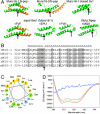
Domain 3a. (A) Domains 3a of structurally characterized SM proteins are shown in green. Munc18-1–Sx4 N peptide and rerefined Munc18-3–Sx4 N peptide reported here are in the Top Left; other structures are indicated by their name and PDB code. For those structures exhibiting a coiled-coil-like interaction, the crystallographically related domain 3a is shown in orange. Arrows indicate the hinge point (equivalent to P335 of Munc18-1) where domain 3a folds back in the rat Munc18-1 structure bound to closed Sx1, showing that other domain 3a structures are more extended. (B) Sequence alignment of domains 3a from Munc18 proteins (rM18-1, rat Munc18-1; hM18-1, human Munc18-1; sM18, squid Sec1; rM18-2, rat Munc18-2; and mM18-3, mouse Munc18-3). Conserved residues are shaded. Helical residues from the structure reported here are indicated by “h.” The conserved proline is indicated by an arrow. (C) Helical wheel representation of helical residues 327–344 of rat Munc18-1, showing hydrophobic residues in yellow, polar residues in green, acidic residues in pink, and basic residues in blue. (D) CD spectra for the domain 3a peptide, residues 326–359 of rat Munc18-1 (blue), SNARE complex (green), and mixture of the peptide and SNARE complex (red). The theoretical curve assuming no interaction between the two is shown in purple.
Fig. 5.
Fig. 5.
Proposed model of Munc18-1–Sx1a interactions. (Left) Closed Sx1 (Habc helices in salmon, SNARE H3 helix in pink) interacts with Munc18-1 (domains 1, 2, and 3 labeled) when the N-peptide binding mode is inactivated; for example, when the N peptide is tagged, Sx1 Ser14 is phosphorylated or the N-peptide binding site of Munc18-1 is blocked. (Center) When the N peptide (N) is engaged on domain 1, Munc18-1 can bind open Sx1 possibly through rotation of domain 1 (yellow/black) relative to domains 2 and 3; this may twist Habc away from H3 and allow extension of the Munc18-1 domain 3a helical hairpin, which we propose binds Sx1 H3. See also Movie S2. (Right) We propose that the extended domain 3a interacts with SNARE complex.

References

    1. Jahn R, Scheller RH. SNAREs—Engines for membrane fusion. Nat Rev Mol Cell Biol. 2006;7:631–643. - PubMed
    1. Südhof T, Rothman J. Membrane fusion: Grappling with SNARE and SM proteins. Science. 2009;323:474–477. - PMC - PubMed
    1. Toonen RF, Verhage M. Munc18-1 in secretion: Lonely Munc joins SNARE team and takes control. Trends Neurosci. 2007;30:564–572. - PubMed
    1. Rizo J, Rosenmund C. Synaptic vesicle fusion. Nat Struct Mol Biol. 2008;15:665–674. - PMC - PubMed
    1. Burkhardt P, Hattendorf DA, Weis WI, Fasshauer D. Munc18a controls SNARE assembly through its interactions with the syntaxin N-peptide. EMBO J. 2008;27:923–933. - PMC - PubMed

Publication types