

Review
doi: 10.2337/db10-0454.
Diabetic retinopathy: targeting vasoregression
Affiliations
- PMID: 21193734
- PMCID: PMC3012202
- DOI: 10.2337/db10-0454
Item in Clipboard
Review
Diabetic retinopathy: targeting vasoregression
Diabetes.
2011 Jan.
No abstract available
Figures

Phenotype of vasoregression in the diabetic retina. In both experimental diabetic rats and diabetic humans, capillary occlusions occur. Nondiabetic (A) and 6-month diabetic rat retina with acellular capillaries (arrows) (B). Nondiabetic (C) and diabetic (D) human retinal digest preparation. Periodic acid-Schiff staining (original magnification ×250).

Concept of physiological angiogenesis. 1) In tip cells, VEGF stimulates DLL4/NOTCH signaling via VEGF-R2, thereby inhibiting tip cell formation and inducing VEGF-R1 expression in the endothelial cells downstream. Astrocyte-derived SDF-1 acts as an additional chemoattractant, activating CXCR4 in tip cells. 2) In stalk cells, predominance of VEGF-R1 and activation of Tie-2 by Ang-2 secreted from the tip cell lead to proliferation and survival. 3) Platelet-derived growth factor receptor (PDGFR)-β+–pericytes are attracted to the growing sprout by PDGF-B, released from tip cells. Interaction of recruited pericytes with endothelial cell–derived Jagged-1 induces the expression of Notch3 and activation of an autoregulatory loop that further enhances Notch3 activation, thereby promoting pericyte survival, investment, vascular branching, and induction of smooth muscle cell (SMC) genes. 4) Transforming growth factor (TGF)-β produced in endothelial cells further induces SMC differentiation and pericytes-derived Ang-1 binds to and activates the Tie-2 receptor on endothelial cells, thereby stimulating vessel maturation and stabilization.

In the mature vasculature, pericyte-derived Ang-1 dominates Ang-2, leading to Tie-2 phosphorylation in endothelial cells. Activation of Tie-2 controls endothelial cell proliferation and induces intercellular contacts and junctions, thereby stabilizing retinal vasculature and promoting the formation of the blood-retinal barrier (A). Diabetes-induced vasoregression is a result of Ang-2 upregulation in the absence of hypoxia. Retinal endothelial cells and glial cells (Müller cells) express Ang-2 as a dominant negative ligand blocking Tie-2 phosphorylation. Upregulation of Ang-2 induces vascular cell depletion and progressive capillary occlusion (B). Growing areas of nonperfusion lead to upregulation of hypoxia-induced factors such as VEGF and Ang-2. In pericytes, Notch3 activation under hypoxic conditions induces Ang-2 expression. Abundance of VEGF without a succinct gradient and elevated Ang-2 levels destabilize vessels, cause endothelial cell proliferation and pericyte activation. Bone marrow–derived progenitor cells contribute to pathological angiogenesis (C). P, phosphate. (A high-quality color representation of this figure is available in the online issue.)

Mechanism of hyperglycemia-induced Ang-2 regulation. A: Under physiological (normoglycemic) conditions, a transcriptional complex (involving the transcriptional corepressor mSin3A) represses Ang-2 transcription by binding to a glucose-sensitive GC box. B: Transcriptional activation of Ang-2 through methylglyoxal (AGE)-induced and hexosamine-propagated modification of SP-3 binding in favor of SP-1 binding. GAPDH, glyceraldehyde-3-phosphate dehydrogenase; UDP, uridine-5-diphosphate. (A high-quality color representation of this figure is available in the online issue.)

Schematic illustration linking hyperglycemia-induced reactive oxygen species (ROS) overproduction with Ang-2–dependent vasoregression and combined ischemia/hypoxia-induced angiogenesis. In healthy retinal capillaries, proper pericyte coverage ensures endothelial cell survival and integrity of blood-retinal barrier by Ang-1/Tie-2 signaling. Chronic hyperglycemia induces cell damage and upregulation of Ang-2 in retinal endothelial cells and Müller cells (MC), leading to retinal pericyte detachment, migration, apoptosis, and progressive vasoregression. Occluded remnants of capillaries are no longer perfused, leading to the upregulation of survival/growth factors such as VEGF. During the later stages, which is not represented in rodent models, increased expression of hypoxia-induced VEGF and increased Ang-2 levels lead to preretinal neovascularization. HXP, hexosamine pathway; EC, endothelial cell. (A high-quality color representation of this figure is available in the online issue.)
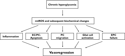
Summary illustration of mechanisms by which chronic hyperglycemia causes intraretinal vasoregression. EC, endothelial cell; EPC, endothelial progenitor cells; mtROS, mitochondrial reactive oxygen species; PC, pericytes.
References
-
- Frank RN: Diabetic retinopathy. N Engl J Med 2004;350:48–58 - PubMed
-
- Gariano RF, Gardner TW: Retinal angiogenesis in development and disease. Nature 2005;438:960–966 - PubMed
-
- Duh E, Aiello LP: Vascular endothelial growth factor and diabetes: the agonist versus antagonist paradox. Diabetes 1999;48:1899–1906 - PubMed
-
- Carmeliet P: Angiogenesis in health and disease. Nat Med 2003;9:653–660 - PubMed
Publication types
MeSH terms
Substances
LinkOut - more resources
Full Text Sources
Medical
