

Contractile response of human anterior vaginal muscularis in women with and without pelvic organ prolapse
- PMID: 21193802
- PMCID: PMC3343064
- DOI: 10.1177/1933719110392054
Contractile response of human anterior vaginal muscularis in women with and without pelvic organ prolapse
Abstract
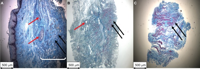
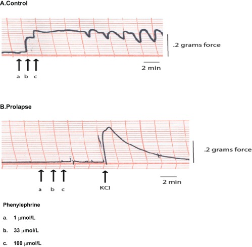
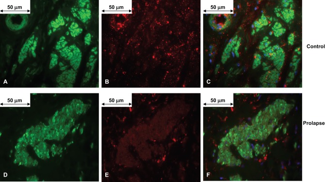
The aim of this study was to compare the contractility of the anterior vaginal muscularis (AVM) from women with and without pelvic organ prolapse (POP). In vitro experiments were performed to measure the peak force generated in response to potassium chloride (KCl; 125 mmol/L) and phenylephrine by AVM tissue from women with and without POP. Cross-sectional areas and co-localization of α(1A) adrenergic receptor protein with smooth muscle α-actin in AVM strips were determined by histology and immunofluorescence, respectively. There were no differences in the mean amplitude of force generated in response to KCl normalized to either wet weight or muscle cross-sectional area (mN/mm(2)) between women with and without POP (P > .30). However, AVM from women with prolapse produced a significantly higher mean force to KCl normalized to total cross-sectional area compared to controls (P = .007). While the control samples demonstrated a consistent response to phenylephrine, there was no response to this stimulant generated by AVM tissue from women with POP. The proportion of co-localized α(1A) adrenergic receptors with smooth muscle α actin in AVM tissue was significantly less in women with POP compared to normal controls (P < .0001). Although there was significantly greater tissue stress generated by AVM from women with prolapse compared to controls, there were no differences in muscle stress. Absent response to phenylephrine by AVM from women with prolapse may be related to a lower expression of α(1A) adrenergic receptors in vaginal smooth muscle.
Conflict of interest statement
The authors declared no potential conflicts of interests with respect to the authorship and/or publication of this article.
Figures
Similar articles
-
The impact of prolapse mesh on vaginal smooth muscle structure and function.BJOG. 2016 Jun;123(7):1076-85. doi: 10.1111/1471-0528.13514. Epub 2015 Aug 20. BJOG. 2016. PMID: 26301457 Free PMC article.
-
Impact of polypropylene prolapse mesh on vaginal smooth muscle in rhesus macaque.Am J Obstet Gynecol. 2019 Oct;221(4):330.e1-330.e9. doi: 10.1016/j.ajog.2019.05.008. Epub 2019 May 16. Am J Obstet Gynecol. 2019. PMID: 31102587 Free PMC article.
-
Changes in elastin density in different locations of the vaginal wall in women with pelvic organ prolapse.Int Urogynecol J. 2014 Dec;25(12):1673-81. doi: 10.1007/s00192-014-2431-9. Epub 2014 May 28. Int Urogynecol J. 2014. PMID: 24866277
-
How to use the Pelvic Organ Prolapse Quantification (POP-Q) system?Neurourol Urodyn. 2018 Aug;37(S6):S39-S43. doi: 10.1002/nau.23740. Neurourol Urodyn. 2018. PMID: 30614056 Review.
-
The role of smooth muscle cells in the pathophysiology of pelvic organ prolapse.Female Pelvic Med Reconstr Surg. 2013 Sep-Oct;19(5):254-9. doi: 10.1097/SPV.0b013e31829ff74d. Female Pelvic Med Reconstr Surg. 2013. PMID: 23982572 Review.
Cited by
-
Extracellular matrix regenerative graft attenuates the negative impact of polypropylene prolapse mesh on vagina in rhesus macaque.Am J Obstet Gynecol. 2017 Feb;216(2):153.e1-153.e9. doi: 10.1016/j.ajog.2016.09.073. Epub 2016 Sep 8. Am J Obstet Gynecol. 2017. PMID: 27615441 Free PMC article.
-
Advances in vaginal bioengineering: Applications, techniques, and needs.Curr Res Physiol. 2023 Oct 18;6:100111. doi: 10.1016/j.crphys.2023.100111. eCollection 2023. Curr Res Physiol. 2023. PMID: 38107786 Free PMC article. Review. No abstract available.
-
In vitro differentiation of endometrial regenerative cells into smooth muscle cells: Α potential approach for the management of pelvic organ prolapse.Int J Mol Med. 2016 Jul;38(1):95-104. doi: 10.3892/ijmm.2016.2593. Epub 2016 May 17. Int J Mol Med. 2016. PMID: 27221348 Free PMC article.
-
Smooth muscle contribution to vaginal viscoelastic response.J Mech Behav Biomed Mater. 2023 Apr;140:105702. doi: 10.1016/j.jmbbm.2023.105702. Epub 2023 Feb 3. J Mech Behav Biomed Mater. 2023. PMID: 36764168 Free PMC article.
-
Vaginal degeneration following implantation of synthetic mesh with increased stiffness.BJOG. 2013 Jan;120(2):233-243. doi: 10.1111/1471-0528.12085. BJOG. 2013. PMID: 23240802 Free PMC article.
References
-
- Olsen AL, Smith VJ, Bergstrom JO, Colling JC, Clark Al. Epidemiology of surgically managed pelvic organ prolapse and urinary incontinence. Obstet Gynecol. 1997;89(4):501–506 - PubMed
-
- Mant J, Painter R, Vessey M. Epidemiology of genital prolapse: observations from the Oxford Family Planning Association Study. Br J Obstet Gynaecol. 1997;104(5):579–585 - PubMed
-
- Smith AR, Hosker GL, Warrell DW. The role of partial denervation of the pelvic floor in the aetiology of genitourinary prolapse and stress incontinence of urine. A neurophysiological study. Br J Obstet Gynaecol. 1989;96(1):24–28 - PubMed
-
- Jackson SR, Avery NC, Tarlton JF, Eckford SD, Abrams P, Bailey AJ. Changes in metabolism of collagen in genitourinary prolapse. Lancet. 1996;347(9016):1658–1661 - PubMed
-
- Ewies AA, Al-Azzawi F, Thompson J. Changes in extracellular matrix proteins in the cardinal ligaments of post-menopausal women with or without prolapse: a computerized immunohistomorphometric analysis. Hum Reprod. 2003;18(10):2189–2195 - PubMed
Publication types
MeSH terms
Substances
Grants and funding
LinkOut - more resources
Full Text Sources
Medical
