

Hippocampal hyperactivation in presymptomatic familial Alzheimer's disease
- PMID: 21194156
- PMCID: PMC3175143
- DOI: 10.1002/ana.22105
Hippocampal hyperactivation in presymptomatic familial Alzheimer's disease
Erratum in
- Ann Neurol. 2011 Jul;70(1):187
Abstract
Objective: The examination of individuals who carry fully penetrant genetic alterations that result in familial Alzheimer's disease (FAD) provides a unique model for studying the early presymptomatic disease stages. In AD, deficits in episodic and associative memory have been linked to structural and functional changes within the hippocampal system. This study used functional MRI (fMRI) to examine hippocampal function in a group of healthy, young, cognitively-intact presymptomatic individuals (average age 33.7 years) who carry the E280A presenilin-1 (PS1) genetic mutation for FAD. These PS1 subjects will go on to develop the first symptoms of the disease around the age of 45 years. Our objective was to examine hippocampal function years before the onset of clinical symptoms.
Methods: Twenty carriers of the Alzheimer's-associated E280A PS1 mutation and 19 PS1-negative control subjects participated. Both groups were matched for age, sex, education level, and neuropsychological test performance. All participants performed a face-name associative encoding task while in a Phillips 1.5T fMRI scanner. Analysis focused on the hippocampal system.
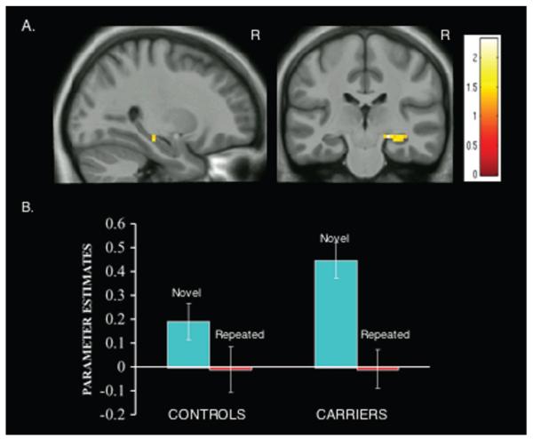
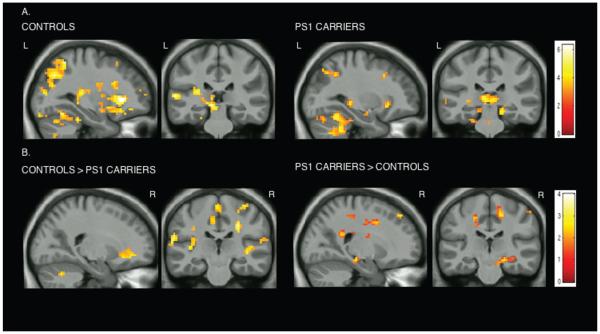
Results: Despite identical behavioral performance, presymptomatic PS1 mutation carriers exhibited increased activation of the right anterior hippocampus during encoding of novel face-name associations compared to matched controls.
Interpretation: Our results demonstrate that functional changes within the hippocampal memory system occur years before cognitive decline in FAD. These presymptomatic changes in hippocampal physiology in FAD suggest that hippocampal fMRI patterns during associative encoding may also provide a preclinical biomarker in sporadic AD.
Figures
References
-
- Alzheimer’s Association . Early onset dementia: a national challenge, a future crisis. Alzheimer’s Association; [Accessed July 18, 2010]. 2006. Available at: http://www.alz.org/national/documents/report_earlyonset_summary.pdf.
-
- Goate A, Chartier-Harlin MC, Mullan M, et al. Segregation of a missense mutation in the amyloid precursor protein gene with familial Alzheimer’s disease. Nature. 1991;349:704–706. - PubMed
-
- Levy-Lahad E, Wasco W, Poorkaj P, et al. Candidate gene for the chromosome 1 familial Alzheimer’s disease locus. Science. 1995;18:973–977. - PubMed
-
- Alzheimer’s Disease Collaborative Group The structure of the presenilin 1 (S182) gene and identification of six novel mutations in early onset AD families. Nat Genet. 1995;11:219–222. - PubMed
-
- Lopera F, Ardilla A, Martínez A, et al. Clinical features of early-onset Alzheimer disease in a large kindred with an E280A presenilin-1 mutation. JAMA. 1997;277:793–799. - PubMed
Publication types
MeSH terms
Substances
Grants and funding
LinkOut - more resources
Full Text Sources
Other Literature Sources
