. 2011 Jun 15;14(12):2327-36.
doi: 10.1089/ars.2010.3526. Epub 2011 Mar 21.

Both maximal expression of selenoproteins and selenoprotein deficiency can promote development of type 2 diabetes-like phenotype in mice

Affiliations

Both maximal expression of selenoproteins and selenoprotein deficiency can promote development of type 2 diabetes-like phenotype in mice

Vyacheslav M Labunskyy et al. Antioxid Redox Signal. .

Abstract

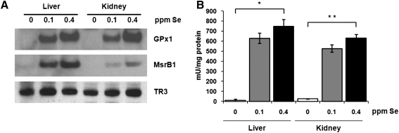
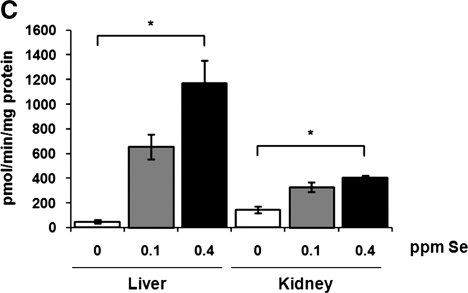
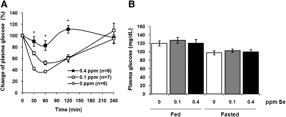
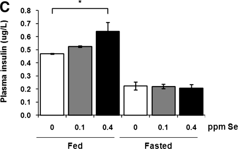
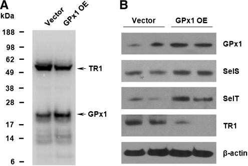
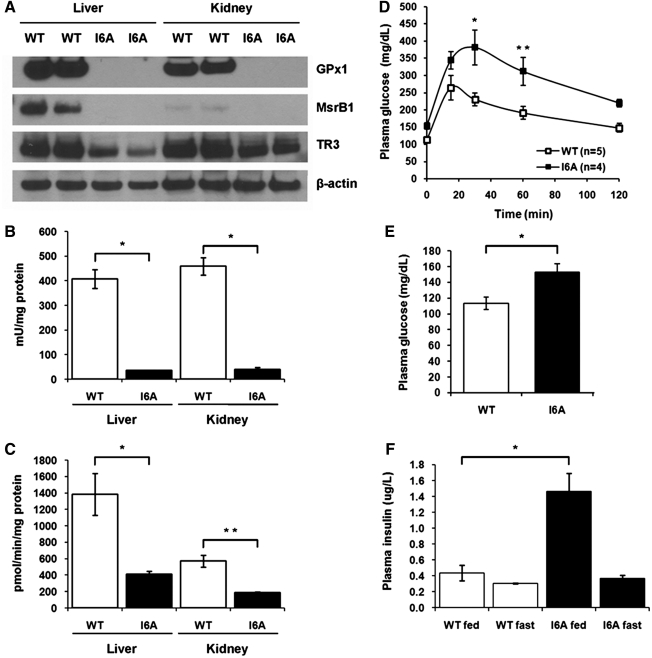
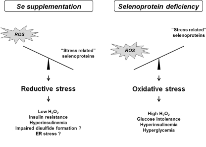
Selenium (Se) is an essential trace element in mammals that has been shown to exert its function through selenoproteins. Whereas optimal levels of Se in the diet have important health benefits, a recent clinical trial has suggested that supplemental intake of Se above the adequate level potentially may raise the risk of type 2 diabetes mellitus. However, the molecular mechanisms for the effect of dietary Se on the development of this disease are not understood. In the present study, we examined the contribution of selenoproteins to increased risk of developing diabetes using animal models. C57BL/6J mice (n=6-7 per group) were fed either Se-deficient Torula yeast-based diet or diets supplemented with 0.1 and 0.4 parts per million Se. Our data show that mice maintained on an Se-supplemented diet develop hyperinsulinemia and have decreased insulin sensitivity. These effects are accompanied by elevated expression of a selective group of selenoproteins. We also observed that reduced synthesis of these selenoproteins caused by overexpression of an i(6)A(-) mutant selenocysteine tRNA promotes glucose intolerance and leads to a diabetes-like phenotype. These findings indicate that both high expression of selenoproteins and selenoprotein deficiency may dysregulate glucose homeostasis and suggest a role for selenoproteins in development of diabetes.


Figures

FIG. 1.
FIG. 1.
Regulation of selenoprotein expression and enzymatic activities by dietary Se in mice. (A) Tissue extracts from C57BL/6J mice maintained on diets containing 0, 0.1, or 0.4 parts per million (ppm) dietary Se were analyzed by SDS-PAGE and Western blotting with antibodies specific for GPx1, MsrB1, and TR3. GPx1 (B) and MsrB (C) activities were analyzed in liver and kidney of mice fed three different Se diets. Results are represented as means±SEM (n=3 for each diet; *p<0.01 and **p<0.001 by two-tailed t-test).
FIG. 1.
FIG. 1.
Regulation of selenoprotein expression and enzymatic activities by dietary Se in mice. (A) Tissue extracts from C57BL/6J mice maintained on diets containing 0, 0.1, or 0.4 parts per million (ppm) dietary Se were analyzed by SDS-PAGE and Western blotting with antibodies specific for GPx1, MsrB1, and TR3. GPx1 (B) and MsrB (C) activities were analyzed in liver and kidney of mice fed three different Se diets. Results are represented as means±SEM (n=3 for each diet; *p<0.01 and **p<0.001 by two-tailed t-test).
FIG. 2.
FIG. 2.
Impaired insulin sensitivity and hyperinsulinemia in mice fed high Se diet. (A) Impact of dietary Se supplementation on insulin sensitivity in C57BL/6J mice. Male C57BL/6J mice maintained on different Se diets for 3 months were fasted overnight, intraperitoneally injected with insulin (0.25 mU/g body weight), and then plasma glucose levels were measured at the indicated time points. Results are expressed as the percentage of the initial glucose levels. Shown are the mean and SEM in each group (n=6–7; *p<0.001 by two-way ANOVA for 0 ppm vs. 0.4 ppm groups). Steady-state plasma glucose (B) and insulin (C) levels were analyzed in mice maintained on different Se diets during fasting (fasted) or in fed state (fed). Results are represented as means±SEM (n=3–10, *p<0.01 by two-tailed t-test).
FIG. 2.
FIG. 2.
Impaired insulin sensitivity and hyperinsulinemia in mice fed high Se diet. (A) Impact of dietary Se supplementation on insulin sensitivity in C57BL/6J mice. Male C57BL/6J mice maintained on different Se diets for 3 months were fasted overnight, intraperitoneally injected with insulin (0.25 mU/g body weight), and then plasma glucose levels were measured at the indicated time points. Results are expressed as the percentage of the initial glucose levels. Shown are the mean and SEM in each group (n=6–7; *p<0.001 by two-way ANOVA for 0 ppm vs. 0.4 ppm groups). Steady-state plasma glucose (B) and insulin (C) levels were analyzed in mice maintained on different Se diets during fasting (fasted) or in fed state (fed). Results are represented as means±SEM (n=3–10, *p<0.01 by two-tailed t-test).
FIG. 3.
FIG. 3.
Effect of GPx1 overexpression on expression levels of other selenoproteins in mice. (A) Liver extracts from wild-type (WT) and GPx1-overexpressing (GPx1 OE) mice, matched for sex and age, were probed by Western blotting with polyclonal antibodies specific for GPx1, MsrB1, SelS, SelT, or monoclonal β-actin-specific antibody as a loading control. Expression of proteins is shown for two WT and two GPx1 OE mice. (B) MsrB activity was analyzed in liver and kidney of WT mice and mice overexpressing GPx1. Results are represented as means±SEM (n=3).
FIG. 4.
FIG. 4.
Contrasting expression patterns of selenoproteins in GPx1-overexpressing and control cell lines. (A), Control cells and cells overexpressing GPx1 were labeled with 75Se, and selenoprotein expression patterns were analyzed by SDS-PAGE followed by autoradiography. Migration of GPx1 and TR1 is shown with arrows. (B) Expression patterns of GPx1, SelS, SelT, and TR1 were analyzed in control and GPx1-overexpressing cell lines by Western blotting. Cell lysates were also probed with monoclonal β-actin-specific antibody as a loading control. The results shown are from two independent experiments.
FIG. 5.
FIG. 5.
Selenoprotein deficiency leads to a dysregulation of glucose homeostasis in i6A mutant Sec tRNA transgenic mice. (A) Tissue extracts from WT or i6A mutant Sec tRNA transgenic mice (I6A) were analyzed by SDS-PAGE and Western blotting with polyclonal antibodies specific for GPx1, MsrB1, TR3, or monoclonal β-actin-specific antibody as a loading control. Expression of proteins is shown for two WT and two I6A transgenic mice. (B) GPx1 activity was analyzed in liver and kidney of WT and I6A transgenic mice. Results are represented as means±SEM (n=3; *p<0.001 by two-tailed t-test). (C) MsrB activity was analyzed in liver and kidney of WT and I6A transgenic mice. Results are represented as means±SEM (n=3; *p<0.05; and **p<0.01 by two-tailed t-test). (D) Effect of an i6A mutant Sec tRNA overexpression on glucose tolerance. 10-month-old WT and I6A males were given a single intraperitoneal injection of glucose (1 mg/g), and plasma glucose levels were measured at the indicated time points. Data are shown as means±SEM in each group (n=4–5; *p<0.01; and **p<0.05 by two-way ANOVA). (E) Fasting plasma glucose levels in WT and I6A mice. Results are represented as means±SEM (n=4–5; *p<0.05 by two-tailed t-test). (F) Steady-state plasma insulin levels in WT and I6A mice during fasting (fast) or in fed state (fed). Results are represented as means±SEM (n=3; *p<0.05 by two-tailed t-test).
FIG. 6.
FIG. 6.
Model for the diabetogenic effect of dietary Se supplementation and selenoprotein deficiency in mice. The effect of supranutritional selenium on the development of diabetes in our Se diet mouse model can be mediated by elevated expression of antioxidant selenoproteins, including GPx1. High GPx1 stimulates reductive stress and prevents normal hydrogen peroxide signaling leading to hyperinsulinemia and decreased insulin sensitivity. However, decreased expression of GPx1 and possibly other selenoproteins in mice overexpressing mutant Sec tRNA may result in high levels of reactive oxygen species (ROS) production and lead to oxidative stress-induced insulin resistance.

References

    1. Behne D. Hilmert H. Scheid S. Gessner H. Elger W. Evidence for specific selenium target tissues and new biologically important selenoproteins. Biochim Biophys Acta. 1988;966:12–21. - PubMed
    1. Bleys J. Navas-Acien A. Guallar E. Selenium and diabetes: more bad news for supplements. Ann Intern Med. 2007;147:271–272. - PubMed
    1. Blot WJ. Li JY. Taylor PR. Guo W. Dawsey S. Wang GQ. Yang CS. Zheng SF. Gail M. Li GY. Nutrition intervention trials in Linxian, China: supplementation with specific vitamin/mineral combinations, cancer incidence, and disease-specific mortality in the general population. J Natl Cancer Inst. 1993;85:1483–1492. - PubMed
    1. Carlson BA. Xu XM. Shrimali R. Sengupta A. Yoo MH. Zhong N. Hatfield DL. Irons R. Davis CD. Lee BJ. Novoselov SV. Gladyshev VN. Mouse models for assessing the role of selenoproteins in health and development. In: Hatfield DL, editor; Berry MJ, editor; Gladyshev VN, editor. Selenium: Its Molecular Biology and Role in Human Health. New York, NY: Springer; 2006. pp. 333–341.
    1. Cheng WH. Ho YS. Ross DA. Han Y. Combs GF., Jr. Lei XG. Overexpression of cellular glutathione peroxidase does not affect expression of plasma glutathione peroxidase or phospholipid hydroperoxide glutathione peroxidase in mice offered diets adequate or deficient in selenium. J Nutr. 1997;127:675–680. - PubMed

Publication types

MeSH terms