Review
. 2011 Sep 15;15(6):1517-30.
doi: 10.1089/ars.2010.3642. Epub 2011 Apr 26.

Regulation of endothelial function by mitochondrial reactive oxygen species

Affiliations
Review

Regulation of endothelial function by mitochondrial reactive oxygen species

Michael E Widlansky et al. Antioxid Redox Signal. .

Abstract

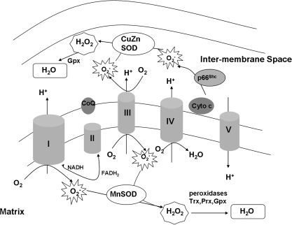
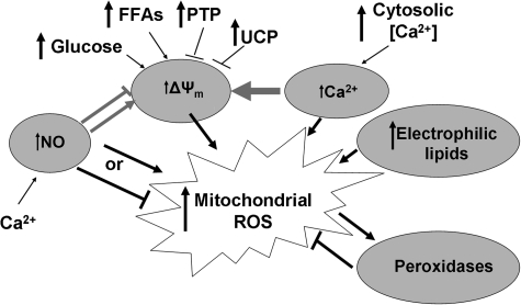
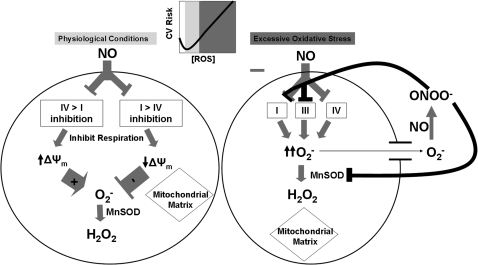
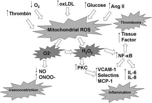
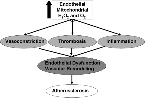
Mitochondria are well known for their central roles in ATP production, calcium homeostasis, and heme and steroid biosynthesis. However, mitochondrial reactive oxygen species (ROS), including superoxide and hydrogen peroxide, once thought to be toxic byproducts of mitochondrial physiologic activities, have recently been recognized as important cell-signaling molecules in the vascular endothelium, where their production, conversion, and destruction are highly regulated. Mitochondrial reactive oxygen species appear to regulate important vascular homeostatic functions under basal conditions in a variety of vascular beds, where, in particular, they contribute to endothelium-dependent vasodilation. On exposure to cardiovascular risk factors, endothelial mitochondria produce excessive ROS in concert with other cellular ROS sources. Mitochondrial ROS, in this setting, act as important signaling molecules activating prothrombotic and proinflammatory pathways in the vascular endothelium, a process that initially manifests itself as endothelial dysfunction and, if persistent, may lead to the development of atherosclerotic plaques. This review concentrates on emerging appreciation of the importance of mitochondrial ROS as cell-signaling molecules in the vascular endothelium under both physiologic and pathophysiologic conditions. Future potential avenues of research in this field also are discussed.


Figures

FIG. 1.
FIG. 1.
Electron-transport chain and reactive oxygen species production. The process of oxidative phosphorylation receives reducing equivalents from the Krebs cycle (NADH to complex I and FADH2 to complex II) and passes these electrons down the transport chain, ultimately to reduce oxygen to water. The fidelity of the process is incomplete, and the relative fidelity of the process depends on local environmental conditions. As such, oxygen can be reduced to O2- in at least three sites within the mitochondria: complexes, I, III, and through p66Shc. O2- that does not escape the mitochondria at this point is rapidly reduced to H2O2 by manganese superoxide dismutase (MnSOD) and copper-zinc superoxide dismutase (CuZnSOD) in the matrix and intermembrane space, respectively. H2O2 either may leave the mitochondria and react with mitochondria proteins, or may be reduced to H2O by local peroxidase enzymes. CoQ, coenzyme Q/ubiquinone; cyto C,cytochrome c.
FIG. 2.
FIG. 2.
Regulation of mitochondrial ROS production. Major regulators of mitochondrial ROS production include nitric oxide (NO), calcium, the mitochondrial membrane potential (ΔΨm), and electrophilic lipids. Interestingly, these effects are often coordinated. ΔΨm is highly influenced by the cellular fuel supply, and uncoupling proteins (UCPs) and the permeability transition pore (PTP) also play significant roles in modulation of mitochondrial ROS through ΔΨm.
FIG. 3.
FIG. 3.
Regulation of mitochondrial ROS in endothelium by NO. The overall effect of NO on mitochondrial ROS production is highly dependent on the relative inhibition of complex IV versus complex I under normal physiologic conditions. Complex IV inhibition occurs rapidly, and complex I inhibition appears to occur over a more prolonged time span. With excessive oxidative stress, as in the setting of cardiovascular risk factors (CV Risk) and atherosclerosis, superoxide reacts rapidly with NO to form peroxynitrite (ONOO-), which inhibits multiple complexes of the respiratory chain as well as MnSOD. MnSOD is overwhelmed, leading to a significant increase in mitochondria-centered superoxide production. Importantly, an optimal level of ROS generation seems to exist, such that both excessive production of ROS (with reduced bioavailability of NO) and insufficient ROS (with attendant impaired physiologic signaling) can lead to a proinflammatory state and promote atherosclerosis.
FIG. 4.
FIG. 4.
Endothelial cell mitochondrial ROS response to pathologic stressors. A wide variety of pathologic stressors associated with cardiovascular risk factors are known to increase mitochondrial ROS production, including thrombin, hypoxemia, oxidized LDL, elevated angiotensin II levels, and elevated glucose levels. These have the effect of increasing both superoxide and hydrogen peroxide levels. This results in inactivation of endothelium-derived NO synthase and consumes any NO that is produced, unleashing proinflammatory signaling pathways. The consequence is increased expression of endothelial cell-adhesion molecules (VCAM-1, selectins, MCP-1), inflammatory cytokines (IL-6 and IL-8), and prothrombotic tissue factor.
FIG. 5.
FIG. 5.
Cascade of risk from excessive endothelial mitochondrial ROS production. Excessive mitochondrial ROS production induces endothelial dysfunction through multiple cell-signaling pathways. The endothelium develops this pathologic phenotype characterized by vasoconstriction, thrombosis, and inflammation. This condition is a precursor to the development of clinically relevant atherosclerosis and predicts future cardiovascular events.

References

    1. Ai L. Rouhanizadeh M. Wu JC. Takabe W. Yu H. Alavi M. Li R. Chu Y. Miller J. Heistad DD. Hsiai TK. Shear stress influences spatial variations in vascular Mn-SOD expression: implication for LDL nitration. Am J Physiol Cell Physiol. 2008;294:C1576–C1585. - PMC - PubMed
    1. Ali MH. Mungai PT. Schumacker PT. Stretch-induced phosphorylation of focal adhesion kinase in endothelial cells: role of mitochondrial oxidants. Am J Physiol Lung Cell Mol Physiol. 2006;291:L38–L45. - PubMed
    1. Ali MH. Pearlstein DP. Mathieu CE. Schumacker PT. Mitochondrial requirement for endothelial responses to cyclic strain: implications for mechanotransduction. Am J Physiol Lung Cell Mol Physiol. 2004;287:L486–L496. - PubMed
    1. Alvarez S. Valdez LB. Zaobornyj T. Boveris A. Oxygen dependence of mitochondrial nitric oxide synthase activity. Biochem Biophys Res Commun. 2003;305:771–775. - PubMed
    1. Anderson S. Bankier AT. Barrell BG. de Bruijn MH. Coulson AR. Drouin J. Eperon IC. Nierlich DP. Roe BA. Sanger F. Schreier PH. Smith AJ. Staden R. Young IG. Sequence and organization of the human mitochondrial genome. Nature. 1981;290:457–465. - PubMed

Publication types

MeSH terms

LinkOut - more resources