. 2011 Jan 3:11:1.
doi: 10.1186/1471-213X-11-1.

Conditional expression of retrovirally delivered anti-MYCN shRNA as an in vitro model system to study neuronal differentiation in MYCN-amplified neuroblastoma

Affiliations

Conditional expression of retrovirally delivered anti-MYCN shRNA as an in vitro model system to study neuronal differentiation in MYCN-amplified neuroblastoma

Jørn R Henriksen et al. BMC Dev Biol. .

Abstract

Background: Neuroblastoma is a childhood cancer derived from immature cells of the sympathetic nervous system. The disease is clinically heterogeneous, ranging from neuronal differentiated benign ganglioneuromas to aggressive metastatic tumours with poor prognosis. Amplification of the MYCN oncogene is a well established poor prognostic factor found in up to 40% of high risk neuroblastomas.Using neuroblastoma cell lines to study neuronal differentiation in vitro is now well established. Several protocols, including exposure to various agents and growth factors, will differentiate neuroblastoma cell lines into neuron-like cells. These cells are characterized by a neuronal morphology with long extensively branched neurites and expression of several neurospecific markers.

Results: In this study we use retrovirally delivered inducible short-hairpin RNA (shRNA) modules to knock down MYCN expression in MYCN-amplified (MNA) neuroblastoma cell lines. By addition of the inducer doxycycline, we show that the Kelly and SK-N-BE(2) neuroblastoma cell lines efficiently differentiate into neuron-like cells with an extensive network of neurites. These cells are further characterized by increased expression of the neuronal differentiation markers NFL and GAP43. In addition, we show that induced expression of retrovirally delivered anti-MYCN shRNA inhibits cell proliferation by increasing the fraction of MNA neuroblastoma cells in the G1 phase of the cell cycle and that the clonogenic growth potential of these cells was also dramatically reduced.

Conclusion: We have developed an efficient MYCN-knockdown in vitro model system to study neuronal differentiation in MNA neuroblastomas.


Figures

Figure 1
Figure 1
Retrovirally delivered inducible shRNA expression in HEK293 cells. TetR-expressing HEK293 cells (HEK293T-Rex) were transduced with retroviruses expressing anti-Luciferase (a-Luc) or scrambled (Scr) shRNAs. The a-Luc shRNA was expressed from a wt-H1 or the inducible H1-2O2 promoter in the absence (-) or presence (+) of 1 μg/ml doxycyclin (dox) for 48 hrs. Error bars indicate SDs.
Figure 2
Figure 2
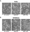
MYCN knockdown induces neurite outgrowth in MNA neuroblastoma cell lines. MNA Kelly (A) and SK-N-BE(2) (B) cells transiently transfected for 3 days with anti-MYCN (aMN-887 and aMN-1658) and scrambled (Scr control) shRNA expressing plasmids. Cells expressing the aMN shRNAs show neurite outgrowth characteristic of neuronal differentiated cells. White arrows indicate neurite outgrowth.
Figure 3
Figure 3
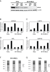
Transient MYCN knockdown in MNA Kelly and SK-N-BE(2) cells using shRNA technology. (A): A representative western blot analysis of MYCN protein expression in Kelly and SK-N-BE(2) cells transiently transfected for 3 days with plasmids expressing the aMN-887 and aMN1658 shRNAs. β-actin expression was used for normalization. Quantitative real-time RT-PCR was used to investigate the expression of MYCN (B), c-MYC (C), neurofilament light-chain - NFL (D) and growth-associated protein 43 - GAP43 (E) mRNAs. Error bars indicate SDs. (F) and (G): Flow cytometric analyses showing the cell cycle distribution of SK-N-BE(2) and Kelly cells transiently transfected with the aMN-887 and aMN-1658 shRNA expressing plasmids. Scr indicates the scrambled shRNA controls.
Figure 4
Figure 4
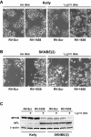
Inducible MYCN knockdown in retrovirally transduced MNA neuroblastoma cell lines. Representative micrographs showing the morphology of Kelly (A) and SK-N-BE(2) (B) cells transduced with retroviruses delivering the dox-inducible aMN-1658 shRNA module (RV-1658). RV-Scr indicates the cells receiving retroviruses delivering an inducible scrambled shRNA module. (C): A representative western blot showing MYCN and NFL expression in Kelly and SK-N-BE(2) cells transduced with retrovirus RV-1658 and induced to express the shRNA by addition of 1 μg/ml doxycyclin (dox).
Figure 5
Figure 5
Induced anti-MYCN shRNA expression decreases MYCN and increases GAP43 expression in MNA neuroblastoma cells. (A) A representative western blot analysis of MYCN, GAP43 and β-actin protein expression in Kelly and SK-N-BE(2) cells induced to express the aMN-1658 shRNA. Real-time RT-PCR analysis of MYCN mRNA (B) and aMN-1658 shRNA (C) expression was performed on total RNA isolated from Kelly and SK-N-BE(2) cells treated as described. Cells were transduced with retrovirus RV-1658 and incubated for the indicated numbers of days in the presence (+) or absence (-) of 1 μg/ml doxycyclin (dox). 3/3 and +/- indicate that the cells were incubated for 3 days in the presence of dox, followed by 3 days in the absence of dox. Error bars indicate SDs.
Figure 6
Figure 6
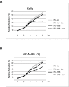
The proliferative inhibition effect of induced MYCN knockdown in MNA neuroblastoma cells. The Alamar Blue Assay was used to measure cell viability in Kelly (A) and SK-N-BE(2) (B) cells transduced with retrovirus RV-Scr (scrambled control) and RV-1658 (anti-MYCN). The addition of doxycyclin (+ dox) induces expression of the shRNAs. AB reduction = Alamar Blue reduction.
Figure 7
Figure 7
Reproductive cell survival in MNA neuroblastoma cells after MYCN knockdown. (A): Graphic presentation of colony forming units (CFU) after induced expression (+ dox) of scrambled (Scr) and anti-MYCN (aMN1658) shRNA in Kelly and SK-N-BE(2) cells. (B): Representative pictures of CFU from retrovirally transduced Kelly cells as presented in A.

References

    1. Maris JM, Hogarty MD, Bagatell R, Cohn SL. Neuroblastoma. Lancet. 2007;369:2106–2120. doi: 10.1016/S0140-6736(07)60983-0. - DOI - PubMed
    1. Bell E, Chen L, Liu T, Marshall GM, Lunec J, Tweddle DA. MYCN oncoprotein targets and their therapeutic potential. Cancer Lett. 2010;293:144–157. doi: 10.1016/j.canlet.2010.01.015. - DOI - PubMed
    1. Kitanaka C, Kato K, Ijiri R, Sakurada K, Tomiyama A, Noguchi K. et al.Increased Ras expression and caspase-independent neuroblastoma cell death: possible mechanism of spontaneous neuroblastoma regression. J Natl Cancer Inst. 2002;94:358–368. - PubMed
    1. Pritchard J, Hickman JA. Why does stage 4 s neuroblastoma regress spontaneously? Lancet. 1994;344:869–870. doi: 10.1016/S0140-6736(94)92834-7. - DOI - PubMed
    1. Bolande RP. The spontaneous regression of neuroblastoma. Experimental evidence for a natural host immunity. Pathol Annu. 1991;26(Pt 2):187–199. - PubMed

Publication types

MeSH terms