. 2011 Jan 28;34(1):122-34.
doi: 10.1016/j.immuni.2010.12.009. Epub 2010 Dec 30.

CD4(+) lymphoid tissue-inducer cells promote innate immunity in the gut

Affiliations

CD4(+) lymphoid tissue-inducer cells promote innate immunity in the gut

Gregory F Sonnenberg et al. Immunity. .

Abstract

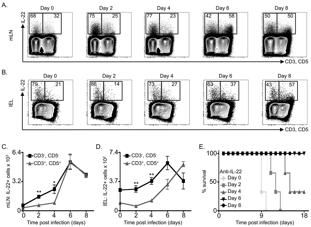
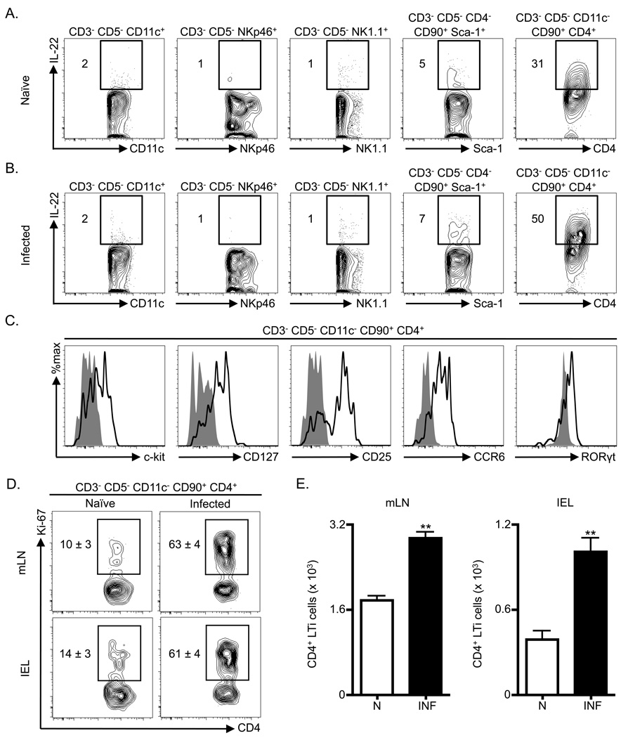
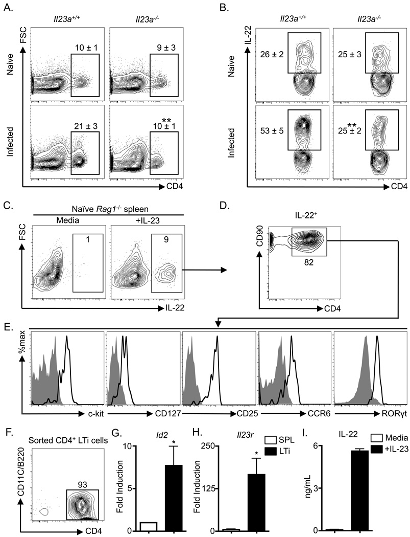
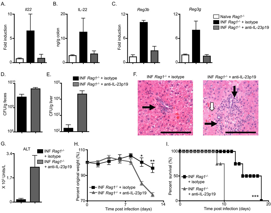
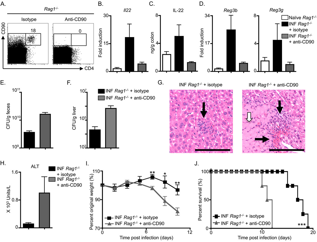
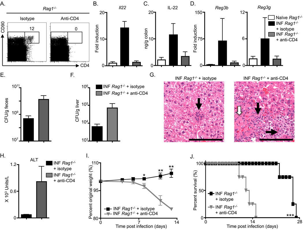
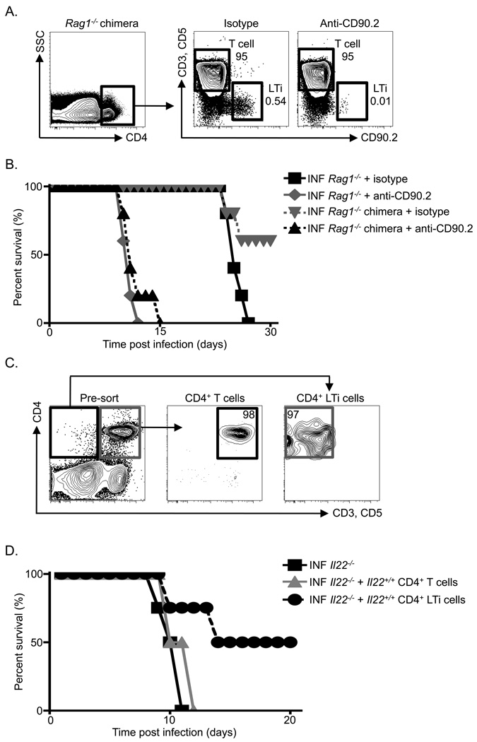
Fetal CD4(+) lymphoid tissue inducer (LTi) cells play a critical role in the development of lymphoid tissues. Recent studies identified that LTi cells persist in adults and are related to a heterogeneous population of innate lymphoid cells that have been implicated in inflammatory responses. However, whether LTi cells contribute to protective immunity remains poorly defined. We demonstrate that after infection with Citrobacter rodentium, CD4(+) LTi cells were a dominant source of interleukin-22 (IL-22) early during infection. Infection-induced CD4(+) LTi cell responses were IL-23 dependent, and ablation of IL-23 impaired innate immunity. Further, depletion of CD4(+) LTi cells abrogated infection-induced expression of IL-22 and antimicrobial peptides, resulting in exacerbated host mortality. LTi cells were also found to be essential for host protective immunity in lymphocyte-replete hosts. Collectively these data demonstrate that adult CD4(+) LTi cells are a critical source of IL-22 and identify a previously unrecognized function for CD4(+) LTi cells in promoting innate immunity in the intestine.


Conflict of interest statement

The authors have no further conflicting financial interests.

Figures

Figure 1
Figure 1. Innate immune cells are the first population to expand with exposure to C. rodentium infection and are the dominant source of IL-22 required for immunity
C57BL/6 mice were infected with C. rodentium on day 0 and sacrificed on days 2, 4, 6 and 8. Cells were briefly stimulated ex vivo and the frequency of IL-22+, CD3+, CD5+ cells versus IL-22+, CD3, CD5 cells were examined in the (A) mLN and (B) colon IEL compartment. Absolute numbers of IL-22+ CD3+/CD5+ cells versus IL-22+, CD3, CD5 cells in the (C) mLN and (D) IEL compartment. (E) Percent survival of IL-22 neutralizing mAb treated and infected C57BL/6 mice. Antibody treatment was initiated on the indicated day and continued every 3 days. All data are representative of 2 independent experiments with a minimum of 3 mice per group or time point. Data shown are the mean ± SEM. * p < 0.05 ** p < 0.01.
Figure 2
Figure 2. Adult CD4+ LTi cells expand and are a dominant innate source of IL-22 following C. rodentium infection
C57BL/6 mice were infected with C. rodentium on day 0 and sacrificed on day 4. Frequency of ex vivo stimulated IL-22+ innate cells in the mLN of (A) naïve and (B) infected mice gated as indicated for various surface markers. (C) The gated CD3 CD5 CD11c CD90+ CD4+ population in the mLN of C. rodentium infected mice was stained with anti- c-kit, CD127, CD25, CCR6 and RORγt antibodies (bold black line) and corresponding isotype and negative control antibodies (solid grey histograms). C57BL/6 mice were infected with C. rodentium on day 0 and sacrificed on day 8. (D) Frequency of Ki-67+ CD4+ LTi cells (CD3 CD5 CD11c CD90+ CD4+) in the mLN and IEL compartment. (E) Absolute numbers of CD4+ LTi cells in the mLN and IEL compartment of naïve (N) and infected (INF) mice. All data are representative of 2 or more independent experiments with a minimum of 3–4 mice per group. Data shown are the mean ± SEM. ** p < 0.01. See Figure S1 for additional data.
Figure 3
Figure 3. Citrobacter rodentium-induced adult CD4+ LTi cell responses are dependent on IL-23
Il23a+/+ and Il23a−/− mice were infected with C. rodentium on day 0 and sacrificed on day 8. (A) Frequency of CD4+ LTi cells in the mLN of naïve and infected mice. Populations are gated on live CD3 CD5 CD90+ CD4+ cells. (B) Frequency of ex vivo stimulated IL-22+ CD4+ LTi cells in the mLN. Populations are gated on live CD3 CD5 CD90+ CD4+ cells. All data are representative of 2 or more independent experiments with a minimum of 3–4 mice per group. Naïve Rag1−/− mouse splenocytes were cultured overnight with or without rIL-23. (C) Frequency of IL-22+ cells with gating on live cells. (D) Frequency of CD90+ CD4+ cells in the IL-22+ gated population of (C). (E) The IL-22+ CD90+ CD4+ population of (D) was stained with c-kit, CD127, CD25, CCR6 and RORγt antibodies (bold black lines) and corresponding isotype and negative control antibodies (solid grey histograms). (F) CD4+ LTi cells were purified from naïve Rag1−/− splenocytes and stained with CD4, CD11c and B220 antibodies. Fold induction of (G) Id2 and (H) Il23r mRNA in purified CD4+ LTi cells relative to total unfractionated Rag1−/− splenocytes (SPL). (I) IL-22 protein in culture supernatants from purified CD4+ LTi cells stimulated in the presence or absence of rIL-23. All in vitro data are representative of 2 or more independent experiments with a minimum of 2–3 replicate wells. Data shown are the mean ± SEM. * p < 0.05 ** p < 0.01. See Figure S2 for additional data.
Figure 4
Figure 4. Innate immunity to Citrobacter rodentium is dependent on IL-23
C57BL/6 Rag1−/− were administered an isotype control mAb or an anti-IL-23p19 mAb starting on day 0, infected with C. rodentium on day 0, and sacrificed on day 10. (A) Fold induction of Il22 transcript in colonic RNA from antibody treated and infected (INF) mice compared to naïve mice. (B) IL-22 protein in the supernatant of colon homogenates and (C) Fold induction of Reg3b and Reg3g transcript in colonic RNA from antibody treated and infected mice compared to naïve mice. C. rodentium CFU in the (D) fecal pellets and (E) liver of antibody treated and infected mice. (F) H&E stained histological sections of the liver of antibody treated and infected mice; inflammatory cell infiltrates (black arrows) and necrotic lesions (white arrow). Scale bar, 100 µm. (G) Serum ALT levels, (H) Percent of original whole body weight and (I) percent survival of antibody treated and infected mice. All data are representative of 3 or more independent experiments with a minimum of 3–4 mice per group. Data shown are the mean ± SEM. * p < 0.05 ** p < 0.01 *** p < 0.001.
Figure 5
Figure 5. Anti-CD90 mAb treatment impairs innate immunity to Citrobacter rodentium
C57BL/6 Rag1−/− were administered an isotype control mAb or an anti-CD90 mAb starting on day 0, infected with C. rodentium on day 0, and sacrificed at day 10. (A) Frequency of CD4+ CD90+ cells in Lin gated splenocytes from antibody treated Rag1−/− mice. (B) Fold induction of Il22 transcript in colonic RNA from antibody treated and infected (INF) mice compared to naïve mice. (C) IL-22 protein in the supernatant of colon homogenates and (D) fold induction of Reg3b and Reg3g transcript in colonic RNA from antibody treated and infected mice compared to naïve mice. C. rodentium CFU in the (E) fecal pellets and (F) liver of antibody treated and infected mice. (G) H&E stained histological sections of the liver of antibody treated and infected mice; inflammatory cell infiltrates (black arrows) and necrotic lesions (white arrow). Scale bar, 100 µm. (H) Serum ALT levels, (I) percent of original whole body weight and (J) percent survival of antibody treated and infected mice. All data are representative of 3 or more independent experiments with a minimum of 3–4 mice per group. Data shown are the mean ± SEM. * p < 0.05 ** p < 0.01 *** p < 0.001. See Figure S3 for additional data.
Figure 6
Figure 6. Adult CD4+ LTi cells mediate innate immunity to Citrobacter rodentium
C57BL/6 Rag1−/− were administered an isotype control mAb or anti-CD4 mAb starting on day 0, infected with C. rodentium on day 0, and sacrificed at day 10. (A) Frequency of CD4+ CD90+ cells in Lin gated splenocytes from antibody treated Rag1−/− mice. (B) Fold induction of Il22 transcript in colonic RNA from antibody treated and infected (INF) mice compared to naïve mice. (C) IL-22 protein in the supernatant of colon homogenates and (D) fold induction of Reg3b and Reg3g transcript in colonic RNA from antibody treated and infected mice compared to naïve mice. C. rodentium CFU in the (E) fecal pellets and (F) liver of antibody treated and infected mice. (G) H&E stained histological sections of the liver of antibody treated and infected mice; inflammatory cell infiltrates (black arrows) and necrotic lesions (white arrow). Scale bar, 100 µm. (H) Serum ALT levels, (I) Percent of original whole body weight and (J) percent survival of antibody treated and infected mice. All data are representative of 3 or more independent experiments with a minimum of 3–4 mice per group. Data shown are the mean ± SEM. * p < 0.05 ** p < 0.01 *** p < 0.001.
Figure 7
Figure 7. Adult CD4+ LTi cells are necessary and sufficient to promote innate immunity to C. rodentium infected lymphocyte-replete hosts
(A) Diagram of the generation of CD90-disparate chimeras with representative flow cytometry plots demonstrating depletion of recipient CD4+ LTi cells but not donor CD4+ T cells in anti-CD90.2 mAb treated chimeras in comparison to isotype mAb treated chimeras. (B) Percent survival of isotype and anti-CD90.2 mAb treated and infected (INF) mice. Displayed data are from two independent experiments with 3 mice per group per experiment. (C) Diagram of innate and adaptive immune cell adoptive transfer approaches utilizing Il22+/+ donors and Il22−/− recipients with representative flow cytometry plots demonstrating purity of transferred populations. (D) Percent survival of antibody treated and infected (INF) mice. Displayed data are from two independent experiments with 2 mice per group per experiment.

References

    1. Bonnet F, Chene G, Thiebaut R, Dupon M, Lawson-Ayayi S, Pellegrin JL, Dabis F, Morlat P. Trends and determinants of severe morbidity in HIV-infected patients: the ANRS CO3 Aquitaine Cohort, 2000–2004. HIV Med. 2007;8:547–554. - PubMed
    1. Buonocore S, Ahern PP, Uhlig HH, Ivanov II, Littman DR, Maloy KJ, Powrie F. Innate lymphoid cells drive interleukin-23-dependent innate intestinal pathology. Nature. 2010;464:1371–1375. - PMC - PubMed
    1. Burnet FM. Evolution of the immune process in vertebrates. Nature. 1968;218:426–430. - PubMed
    1. Cella M, Fuchs A, Vermi W, Facchetti F, Otero K, Lennerz JK, Doherty JM, Mills JC, Colonna M. A human natural killer cell subset provides an innate source of IL-22 for mucosal immunity. Nature. 2009;457:722–725. - PMC - PubMed
    1. Colonna M. Interleukin-22-producing natural killer cells and lymphoid tissue inducer-like cells in mucosal immunity. Immunity. 2009;31:15–23. - PubMed

Publication types

MeSH terms