

Direct inhibition of hypocretin/orexin neurons in the lateral hypothalamus by nociceptin/orphanin FQ blocks stress-induced analgesia in rats
- PMID: 21195099
- PMCID: PMC3031765
- DOI: 10.1016/j.neuropharm.2010.12.026
Direct inhibition of hypocretin/orexin neurons in the lateral hypothalamus by nociceptin/orphanin FQ blocks stress-induced analgesia in rats
Abstract
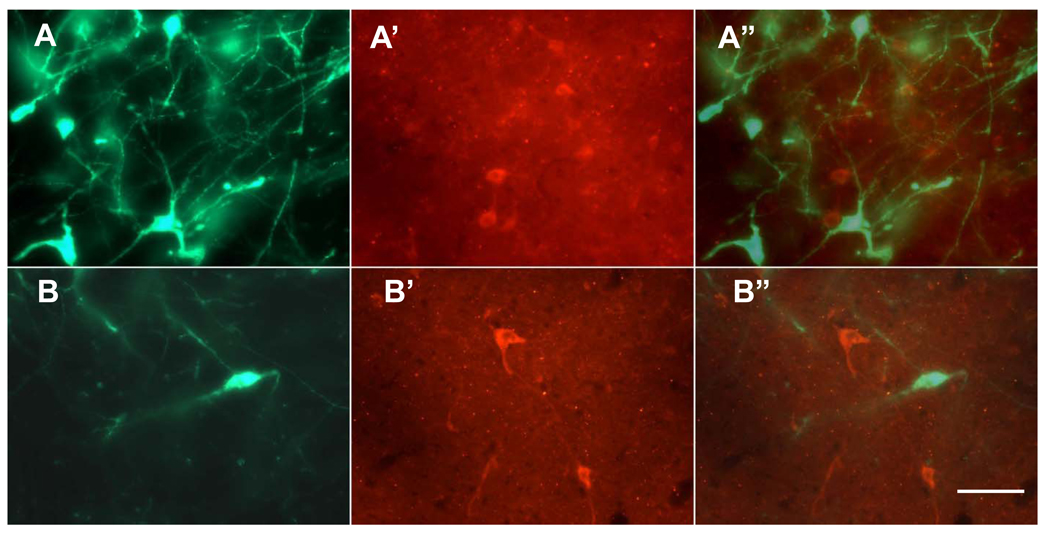
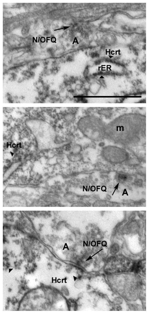
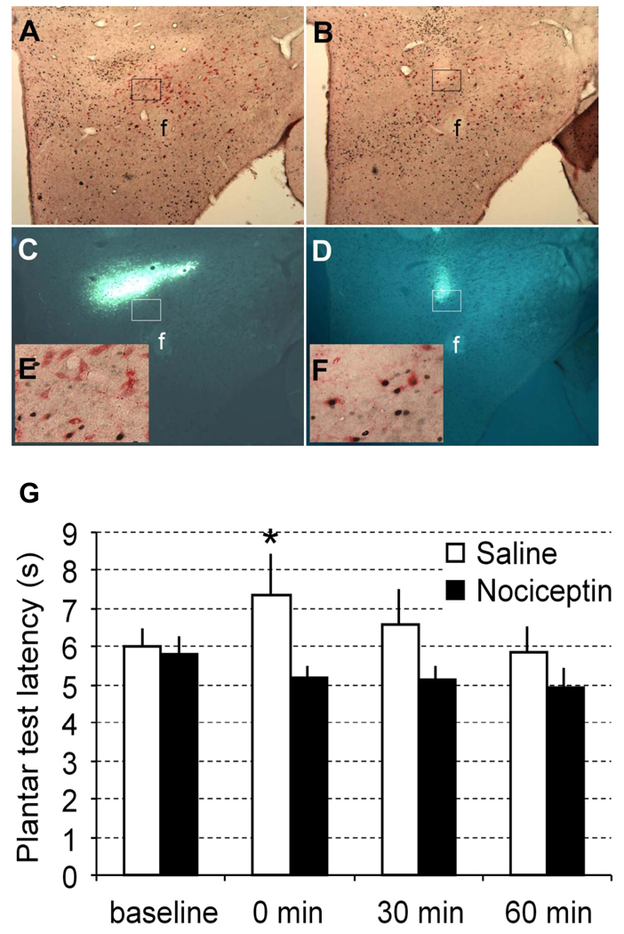
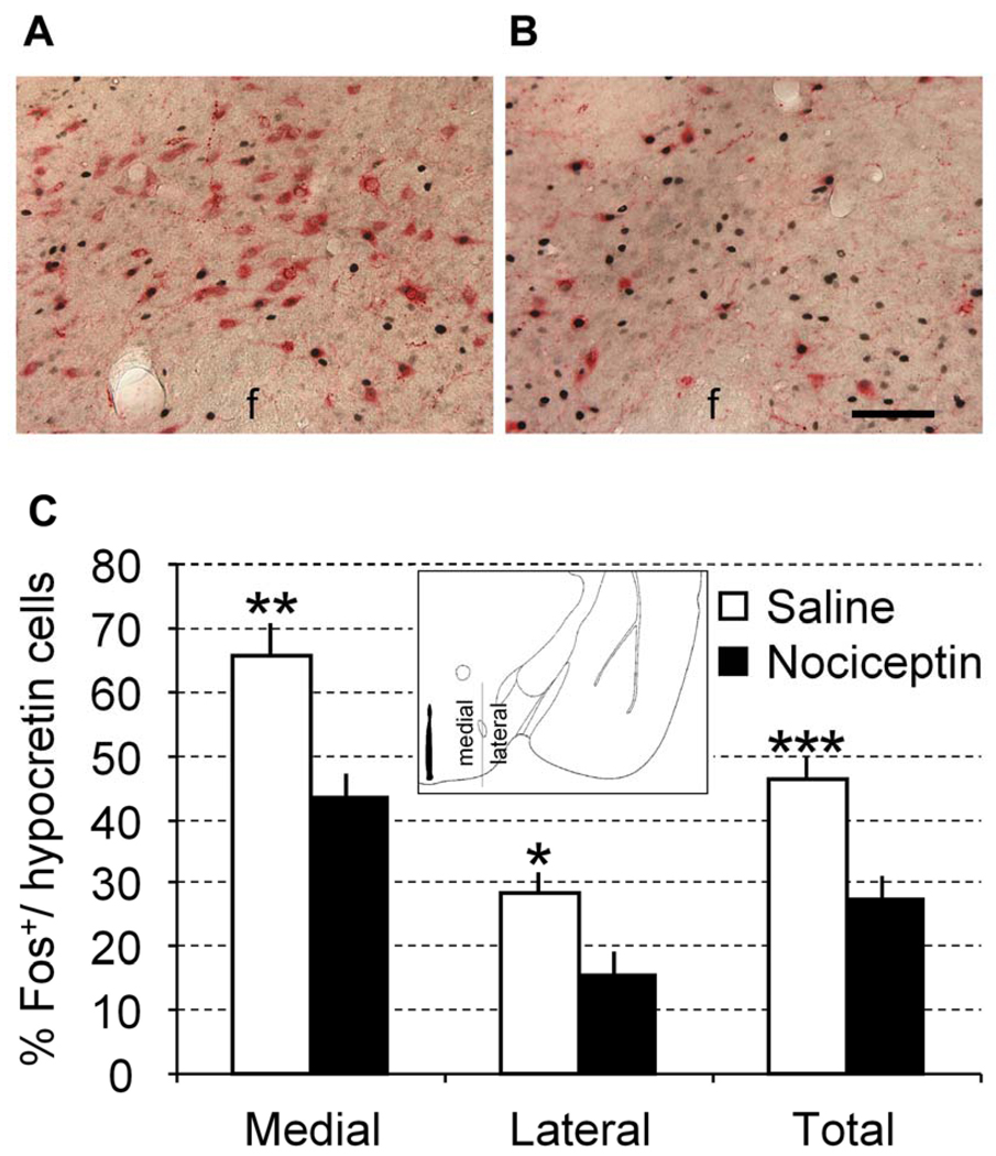
We recently demonstrated that hypocretin/orexin (Hcrt) and nociceptin/orphanin FQ (N/OFQ) systems coordinately regulate nociception in a mouse model of stress-induced analgesia (SIA). However, the site of N/OFQ action on modulation of SIA was elusive, since N/OFQ was administered via intracerebroventricular (i.c.v.) injection acting on widely distributed N/OFQ receptors (NOP) in the brain. In the present study, we tested the hypothesis that N/OFQ modulates the SIA directly via the inhibition of the Hcrt neurons in the lateral hypothalamus. Using both fluorescent and electron microscopy, we found that N/OFQ-containing neurons are located in the lateral hypothalamus and the N/OFQ-containing fibers make direct contacts with the Hcrt neurons. Paw thermal nociceptive test revealed that the immobilization restraint of the rat increased the thermal pain threshold by 20.5 ± 7.6%. Bilateral microinjection of N/OFQ (9 μg/side) into the rat perifornical area of the lateral hypothalamus, the brain area in which the Hcrt neurons are exclusively located, abolished the SIA. Activity of Hcrt neurons in the same animals was assessed using Fos immunohistochemistry. Percentage of Fos(+)/Hcrt neurons was lower in rats injected with N/OFQ than rats injected with saline, with the difference between groups stronger in the Hcrt neurons located medially to the fornix than in Hcrt neurons located laterally to the fornix. These results suggest that N/OFQ modulation of SIA is mediated by direct inhibition of Hcrt neuronal activity in the perifornical area. The uncovered peptidergic interaction circuitry may have broad implication in coordinated modulation by Hcrt and N/OFQ on other stress adaptive responses.
Copyright © 2010 Elsevier Ltd. All rights reserved.
Figures
Similar articles
-
Hypocretin/orexin and nociceptin/orphanin FQ coordinately regulate analgesia in a mouse model of stress-induced analgesia.J Clin Invest. 2008 Jul;118(7):2471-81. doi: 10.1172/JCI35115. J Clin Invest. 2008. PMID: 18551194 Free PMC article.
-
The neuronal circuit between nociceptin/orphanin FQ and hypocretins/orexins coordinately modulates stress-induced analgesia and anxiety-related behavior.Vitam Horm. 2015;97:295-321. doi: 10.1016/bs.vh.2014.11.004. Epub 2015 Jan 20. Vitam Horm. 2015. PMID: 25677777 Review.
-
Nociceptin induces hypophagia in the perifornical and lateral hypothalamic area.PLoS One. 2012;7(9):e45350. doi: 10.1371/journal.pone.0045350. Epub 2012 Sep 17. PLoS One. 2012. PMID: 23028954 Free PMC article.
-
Co-localization of hypocretin-1 and leucine-enkephalin in hypothalamic neurons projecting to the nucleus of the solitary tract and their effect on arterial pressure.Neuroscience. 2013 Oct 10;250:599-613. doi: 10.1016/j.neuroscience.2013.07.054. Epub 2013 Jul 30. Neuroscience. 2013. PMID: 23912034
-
The biology of Nociceptin/Orphanin FQ (N/OFQ) related to obesity, stress, anxiety, mood, and drug dependence.Pharmacol Ther. 2014 Mar;141(3):283-99. doi: 10.1016/j.pharmthera.2013.10.011. Epub 2013 Nov 1. Pharmacol Ther. 2014. PMID: 24189487 Free PMC article. Review.
Cited by
-
Differences in carbachol dose, pain condition, and sex following lateral hypothalamic stimulation.Neuroscience. 2014 Jun 13;270:226-35. doi: 10.1016/j.neuroscience.2014.04.020. Epub 2014 Apr 20. Neuroscience. 2014. PMID: 24759771 Free PMC article.
-
Odour-induced analgesia mediated by hypothalamic orexin neurons in mice.Sci Rep. 2016 Nov 15;6:37129. doi: 10.1038/srep37129. Sci Rep. 2016. PMID: 27845440 Free PMC article.
-
Neuropeptide S-initiated sequential cascade mediated by OX1, NK1, mGlu5 and CB1 receptors: a pivotal role in stress-induced analgesia.J Biomed Sci. 2020 Jan 9;27(1):7. doi: 10.1186/s12929-019-0590-1. J Biomed Sci. 2020. PMID: 31915019 Free PMC article.
-
Orexin A and orexin receptor 1 axonal traffic in dorsal roots at the CNS/PNS interface.Front Neurosci. 2014 Feb 11;8:20. doi: 10.3389/fnins.2014.00020. eCollection 2014. Front Neurosci. 2014. PMID: 24574957 Free PMC article.
-
A Bitter Experience That Enlightens the Future: COVID-19 Neurological Affection and Perspectives on the Orexigenic System.Cureus. 2022 Oct 28;14(10):e30788. doi: 10.7759/cureus.30788. eCollection 2022 Oct. Cureus. 2022. PMID: 36457603 Free PMC article. Review.
References
-
- Amit Z, Galina ZH. Stress-induced analgesia: adaptive pain suppression. Physiol Rev. 1986;66:1091–1120. - PubMed
-
- Amit Z, Galina ZH. Stress induced analgesia plays an adaptive role in the organization of behavioral responding. Brain Res.Bull. 1988;21:955–958. - PubMed
-
- Anton B, Fein J, To T, Li X, Silberstein L, Evans CJ. Immunohistochemical localization of ORL-1 in the central nervous system of the rat. J.Comp Neurol. 1996 Apr 29;368:229–251. - PubMed
-
- Blair R, Galina ZH, Holmes LJ, Amit Z. Stress-induced analgesia: A performance deficit or a change in pain responsiveness? Behav.Neural Biol. 1982;34:152–158. - PubMed
-
- Butler RK, Finn DP. Stress-induced analgesia. Prog.Neurobiol. 2009;88:184–202. - PubMed
Publication types
MeSH terms
Substances
Grants and funding
LinkOut - more resources
Full Text Sources
Medical
