Review
. 2011 Jun;48(11):1292-300.
doi: 10.1016/j.molimm.2010.12.006. Epub 2010 Dec 31.

L2pB1: a new player in autoimmunity

Affiliations
Review

L2pB1: a new player in autoimmunity

Xuemei Zhong et al. Mol Immunol. 2011 Jun.

Abstract

L2pB1 cells (PD-L2positive B1 cells) are a newly discovered subpopulation of B1 B cells. L2pB1 cells are noted for the expression of PD-L2 (CD273, B7-DC), a ligand for the inhibitory receptor PD-1, which distinguishes this subpopulation from other B1 B cells that lack PD-L2, namely, L2nB1 cells (PD-L2negative B1 cells). PD-L2 gene expression is regulated differently in B1 B cells as compared to macrophages and dendritic cells. L2pB1 cells share many commonly known B1 cell features with L2nB1 cells. These include spontaneous IgM secretion, constitutive ERK activation, elevated co-stimulatory molecule expression, skewing of T cell differentiation, and unique proliferative responsiveness (to LPS, PMA, but not anti-IgM). However, L2pB1 cells express a biased Ig repertoire that is enriched for self-reactivity as compared with L2nB1 cells. Further, L2pB1 cells present antigen more potently than L2nB1 cells. In addition, L2pB1 cells switch Ig isotype more readily from IgM to IgG1 and IgG2b upon cytokine stimulation. Moreover, increased numbers of L2pB1 cells are present in murine models of lupus and this correlates with increased serum anti-dsDNA titers. These characteristics suggest that L2pB1 cells may play a pathophysiological role in autoimmune dyscrasias. In this report we review the special features of L2pB1 cells and how they may contribute to autoimmunity.


Figures

Figure 1
Figure 1. L2pB1 cells are a major B1 B cell population in the peritoneal cavity
(A) Peritoneal washout cells from BALB/c mice were stained with anti-B220 (clone RA3-6B2, eBioscience), anti-CD5 (clone 53-7.3, eBioscience) and anti-PD-L2 (clone TY25, eBioscience) and analyzed by flow cytometry. PD-L2 expression on each cell population is shown. L2pB1 cells and L2nB1 cells are highlighted by arrows. Isotype matched control antibody staining is shown as blue in the histograms. (B) A smear of peritoneal washout cells from BXSB mice was stained with anti-CD20 (clone L26, BioCare), anti-PD-L2 (clone TY25, eBioscience) and DAPI, as indicated, and examined by fluorescence microscopy.
Figure 2
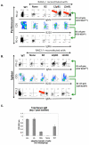
Figure 2. Homing of L2pB1 cells after adoptive transfer into RAG2−/−γc−/− mice
Donor B cells (B2 B cells, L2pB1 cells, L2nB1 cells) were sort-purified from BALB/c mice. C57BL/6J-gCKO-Rag2KO (RAG2−/−) mice were used as recipients. Each recipient mouse received 8 × 10⁵ donor B cells through i.v. injection. On day 13 post-transfer, cells were isolated from recipient mice and analyzed by flow cytometry. Donor B cells were identified as IgMa+ B cells and recipient B cells were IgMa-. RAG2−/− mice received no donor B cells were used as negative controls. Wild type C57BL/6J mice (WT) were used as positive controls for proper gating of each cell population. (A) Peritoneal washout cells from recipient mice were stained for B220 and CD5. B1 and B2 cells were gated as B220lowCD5+ and B220hiCD5 populations respectively (middle panel). After i.v. injection, donor-derived L2pB1 cells (PD-L2+IgMa+) and L2nB1 cells (PD-L2-IgMa+) were found mostly within the peritoneal B1 cell gate (red arrows). No donor B2 B cells were found in the peritoneum after transfer. (B) Total splenocytes from recipient mice were stained for B220 and CD5. B1 and B2 cells were gated as described above (middle panel). B2 B cells were found mostly in the spleen (red arrow) after transfer. Few L2pB1 and L2nB1 were found in the spleen. (C) Total serum IgM from recipients transferred with various donor B cells (L2+: L2pB1 cells, L2-: L2nB1 cells, B2s: splenic B2 cells) and controls (None: C57BL/6J-gCKO-Rag2KO mice received no donor cells, WT: C57BL/6J wild type mice) were analyzed by ELISA. Mean values of three mice in each group are shown, along with lines indicating standard errors of the means.
Figure 3
Figure 3. Potential roles of L2pB1 cells in autoimmunity
Immunity and autoimmunity may represent two sides of the same L2pB1 coin. On one hand, L2pB1 cells can be immune-protective and regulatory. Natural, autoreactive IgM secreted by L2pB1 cells can mask or dispose of self-antigens and thereby prevent immune sensitization and production of pathogenic IgG antibodies. Further, L2pB1 cells can modulate incipient autoimmune responses through engagement of PD-1, expression of FasL, and secretion of IL-10. Conversely, L2pB1 cells can be pathogenic. Broadly reactive IgM secreted by L2pB1 cells provides a first line of defense in acute viral and bacterial infection. However, chronic infection may over-activate L2pB1 cells and provide an opportunity for class-switching and affinity maturation which consequently leads to autoimmunity. L2pB1 cells may stimulate effector T cells and influence naïve T cells to differentiate into pro-inflammatory Th17 and Th1 cells. Thus, whether L2pB1 cells are protective or pathogenic may depend on environmental factors that arise during infection and inflammation.

References

    1. Amel Kashipaz MR, Huggins ML, Lanyon P, Robins A, Powell RJ, Todd I. Assessment of Be1 and Be2 cells in systemic lupus erythematosus indicates elevated interleukin-10 producing CD5+ B cells. Lupus. 2003;12:356–63. - PubMed
    1. Ansel KM, Harris RB, Cyster JG. CXCL13 is required for B1 cell homing, natural antibody production, and body cavity immunity. Immunity. 2002;16:67–76. - PubMed
    1. Baudino L, Nimmerjahn F, Azeredo da Silveira S, Martinez-Soria E, Saito T, Carroll M, Ravetch JV, Verbeek JS, Izui S. Differential contribution of three activating IgG Fc receptors (FcgammaRI, FcgammaRIII, and FcgammaRIV) to IgG2a-and IgG2b-induced autoimmune hemolytic anemia in mice. J Immunol. 2008;180:1948–53. - PubMed
    1. Baumgarth N, Herman OC, Jager GC, Brown LE, Herzenberg LA, Chen J. B-1 and B-2 cell-derived immunoglobulin M antibodies are nonredundant components of the protective response to influenza virus infection. J Exp Med. 2000;192:271–80. - PMC - PubMed
    1. Becker H, Weber C, Storch S, Federlin K. Relationship between CD5+ B lymphocytes and the activity of systemic autoimmunity. Clin Immunol Immunopathol. 1990;56:219–25. - PubMed

Publication types